National Institutes of Health/National Institute of General Medical Sciences (NIH/NIGMS)
U24 GM116789
United States
Citation
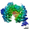
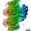
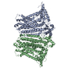
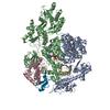
Journal: Proc Natl Acad Sci U S A / Year: 2019 Title: Cryo-EM structure of OSCA1.2 from elucidates the mechanical basis of potential membrane hyperosmolality gating. Authors: Koustav Maity / John M Heumann / Aaron P McGrath / Noah J Kopcho / Po-Kai Hsu / Chang-Wook Lee / James H Mapes / Denisse Garza / Srinivasan Krishnan / Garry P Morgan / Kevin J Hendargo / ...Authors: Koustav Maity / John M Heumann / Aaron P McGrath / Noah J Kopcho / Po-Kai Hsu / Chang-Wook Lee / James H Mapes / Denisse Garza / Srinivasan Krishnan / Garry P Morgan / Kevin J Hendargo / Thomas Klose / Steven D Rees / Arturo Medrano-Soto / Milton H Saier / Miguel Piñeros / Elizabeth A Komives / Julian I Schroeder / Geoffrey Chang / Michael H B Stowell / Abstract: Sensing and responding to environmental water deficiency and osmotic stresses are essential for the growth, development, and survival of plants. Recently, an osmolality-sensing ion channel called ...Sensing and responding to environmental water deficiency and osmotic stresses are essential for the growth, development, and survival of plants. Recently, an osmolality-sensing ion channel called OSCA1 was discovered that functions in sensing hyperosmolality in Here, we report the cryo-electron microscopy (cryo-EM) structure and function of an OSCA1 homolog from rice (; OsOSCA1.2), leading to a model of how it could mediate hyperosmolality sensing and transport pathway gating. The structure reveals a dimer; the molecular architecture of each subunit consists of 11 transmembrane (TM) helices and a cytosolic soluble domain that has homology to RNA recognition proteins. The TM domain is structurally related to the TMEM16 family of calcium-dependent ion channels and lipid scramblases. The cytosolic soluble domain possesses a distinct structural feature in the form of extended intracellular helical arms that are parallel to the plasma membrane. These helical arms are well positioned to potentially sense lateral tension on the inner leaflet of the lipid bilayer caused by changes in turgor pressure. Computational dynamic analysis suggests how this domain couples to the TM portion of the molecule to open a transport pathway. Hydrogen/deuterium exchange mass spectrometry (HDXMS) experimentally confirms the conformational dynamics of these coupled domains. These studies provide a framework to understand the structural basis of proposed hyperosmolality sensing in a staple crop plant, extend our knowledge of the anoctamin superfamily important for plants and fungi, and provide a structural mechanism for potentially translating membrane stress to transport regulation.
Average exposure time: 1 sec. / Electron dose: 55 e/Å2 / Detector mode: SUPER-RESOLUTION / Film or detector model: GATAN K2 SUMMIT (4k x 4k) / Num. of grids imaged: 5 / Num. of real images: 2408
EM imaging optics
Energyfilter name: GIF Bioquantum / Energyfilter slit width: 20 eV / Phase plate: VOLTA PHASE PLATE
Image scans
Movie frames/image: 40 / Used frames/image: 1-40
-
Processing
EM software
ID
Name
Version
Category
1
RELION
2
particleselection
2
Leginon
imageacquisition
7
PHENIX
modelfitting
9
PHENIX
modelrefinement
10
cryoSPARC
initialEulerassignment
11
RELION
finalEulerassignment
CTF correction
Type: PHASE FLIPPING AND AMPLITUDE CORRECTION
Particle selection
Num. of particles selected: 169655
Symmetry
Point symmetry: C2 (2 fold cyclic)
3D reconstruction
Resolution: 4.9 Å / Resolution method: FSC 0.143 CUT-OFF / Num. of particles: 64096 / Num. of class averages: 1 / Symmetry type: POINT
Atomic model building
B value: 400 / Protocol: AB INITIO MODEL / Space: REAL
+
About Yorodumi
-
News
-
Feb 9, 2022. New format data for meta-information of EMDB entries
New format data for meta-information of EMDB entries
Version 3 of the EMDB header file is now the official format.
The previous official version 1.9 will be removed from the archive.
In the structure databanks used in Yorodumi, some data are registered as the other names, "COVID-19 virus" and "2019-nCoV". Here are the details of the virus and the list of structure data.
Jan 31, 2019. EMDB accession codes are about to change! (news from PDBe EMDB page)
EMDB accession codes are about to change! (news from PDBe EMDB page)
The allocation of 4 digits for EMDB accession codes will soon come to an end. Whilst these codes will remain in use, new EMDB accession codes will include an additional digit and will expand incrementally as the available range of codes is exhausted. The current 4-digit format prefixed with “EMD-” (i.e. EMD-XXXX) will advance to a 5-digit format (i.e. EMD-XXXXX), and so on. It is currently estimated that the 4-digit codes will be depleted around Spring 2019, at which point the 5-digit format will come into force.
The EM Navigator/Yorodumi systems omit the EMD- prefix.
Related info.:Q: What is EMD? / ID/Accession-code notation in Yorodumi/EM Navigator
Yorodumi is a browser for structure data from EMDB, PDB, SASBDB, etc.
This page is also the successor to EM Navigator detail page, and also detail information page/front-end page for Omokage search.
The word "yorodu" (or yorozu) is an old Japanese word meaning "ten thousand". "mi" (miru) is to see.
Related info.:EMDB / PDB / SASBDB / Comparison of 3 databanks / Yorodumi Search / Aug 31, 2016. New EM Navigator & Yorodumi / Yorodumi Papers / Jmol/JSmol / Function and homology information / Changes in new EM Navigator and Yorodumi
 Movie
Movie Controller


 Open data
 Basic information
 Components
 Keywords
 TRANSPORT PROTEIN / ion channel osmolality gated
 Function and homology information
 Authors
 United States, 2items
  Citation
 Structure visualization
 Downloads & links
 Other downloads
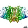


 PDBj
PDBj Assembly
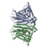

 Sample preparation
 Electron microscopy imaging
 Processing