 Movie
Movie Controller

[English] 日本語
 Yorodumi
- PDB-6o3j: Crystal structure of the Fab fragment of the human HIV-1 neutrali... -
+ Open data

- Basic information
| Entry | Database: PDB / ID: 6o3j | ||||||
|---|---|---|---|---|---|---|---|
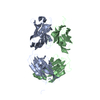
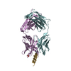
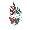
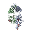
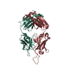
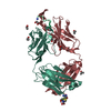
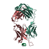
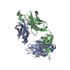
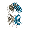
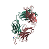
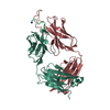
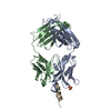
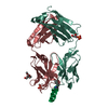
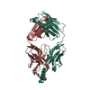
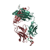
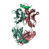
| Title | Crystal structure of the Fab fragment of the human HIV-1 neutralizing antibody PGZL1 in complex with its MPER peptide epitope (region 671-683 of HIV-1 gp41) and phosphatidic acid (06:0 PA) | ||||||
 Components Components |
| ||||||
 Keywords Keywords |  IMMUNE SYSTEM / PGZL1 ANTI HIV-1 / GP41 MPER / MEMBRANE LIPIDS / BROADLY NEUTRALISING HIV-1 ANTIBODY IMMUNE SYSTEM / PGZL1 ANTI HIV-1 / GP41 MPER / MEMBRANE LIPIDS / BROADLY NEUTRALISING HIV-1 ANTIBODY | ||||||
| Function / homology |  Function and homology information Function and homology informationSynthesis and processing of ENV and VPU / evasion of host immune response / Alpha-defensins / Dectin-2 family / Binding and entry of HIV virion / positive regulation of plasma membrane raft polarization / positive regulation of receptor clustering / positive regulation of establishment of T cell polarity / virus-mediated perturbation of host defense response / host cell endosome membrane ...Synthesis and processing of ENV and VPU / evasion of host immune response / Alpha-defensins / Dectin-2 family / Binding and entry of HIV virion / positive regulation of plasma membrane raft polarization / positive regulation of receptor clustering / positive regulation of establishment of T cell polarity / virus-mediated perturbation of host defense response / host cell endosome membrane / actin filament organization / Assembly Of The HIV Virion / Budding and maturation of HIV virion / clathrin-dependent endocytosis of virus by host cell / viral protein processing / symbiont entry into host cell / fusion of virus membrane with host plasma membrane / fusion of virus membrane with host endosome membrane / viral envelope / virion attachment to host cell / host cell plasma membrane / virion membrane / structural molecule activity / membraneSimilarity search - Function | ||||||
| Biological species |  Homo sapiens (human) Homo sapiens (human)  Human immunodeficiency virus 1 Human immunodeficiency virus 1 | ||||||
| Method | X-RAY DIFFRACTION / SYNCHROTRON / MOLECULAR REPLACEMENT / Resolution: 3.416 Å | ||||||
 Authors Authors | Irimia, A. / Wilson, I.A. | ||||||
| Funding support |  United States, 1items United States, 1items
| ||||||
 Citation Citation | Journal: Nat Commun / Year: 2019 Title: An MPER antibody neutralizes HIV-1 using germline features shared among donors. Authors: Lei Zhang / Adriana Irimia / Lingling He / Elise Landais / Kimmo Rantalainen / Daniel P Leaman / Thomas Vollbrecht / Armando Stano / Daniel I Sands / Arthur S Kim / / Pascal Poignard / ...Authors: Lei Zhang / Adriana Irimia / Lingling He / Elise Landais / Kimmo Rantalainen / Daniel P Leaman / Thomas Vollbrecht / Armando Stano / Daniel I Sands / Arthur S Kim / / Pascal Poignard / Dennis R Burton / Ben Murrell / Andrew B Ward / Jiang Zhu / Ian A Wilson / Michael B Zwick /   Abstract: The membrane-proximal external region (MPER) of HIV-1 envelope glycoprotein (Env) can be targeted by neutralizing antibodies of exceptional breadth. MPER antibodies usually have long, hydrophobic ...The membrane-proximal external region (MPER) of HIV-1 envelope glycoprotein (Env) can be targeted by neutralizing antibodies of exceptional breadth. MPER antibodies usually have long, hydrophobic CDRH3s, lack activity as inferred germline precursors, are often from the minor IgG3 subclass, and some are polyreactive, such as 4E10. Here we describe an MPER broadly neutralizing antibody from the major IgG1 subclass, PGZL1, which shares germline V/D-region genes with 4E10, has a shorter CDRH3, and is less polyreactive. A recombinant sublineage variant pan-neutralizes a 130-isolate panel at 1.4 μg/ml (IC). Notably, a germline revertant with mature CDR3s neutralizes 12% of viruses and still binds MPER after DJ reversion. Crystal structures of lipid-bound PGZL1 variants and cryo-EM reconstruction of an Env-PGZL1 complex reveal how these antibodies recognize MPER and viral membrane. Discovery of common genetic and structural elements among MPER antibodies from different patients suggests that such antibodies could be elicited using carefully designed immunogens. | ||||||
| History |
|
- Structure visualization
| Structure viewer | Molecule: MolmilJmol/JSmol |
|---|
- Downloads & links
-Download
| PDBx/mmCIF format | 6o3j.cif.gz | 185.2 KB | Display | PDBx/mmCIF format |
|---|---|---|---|---|
| PDB format | pdb6o3j.ent.gz | 145.1 KB | Display | PDB format |
| PDBx/mmJSON format | 6o3j.json.gz | Tree view | PDBx/mmJSON format | |
| Others |  Other downloads Other downloads |
-Validation report
| Arichive directory | https://data.pdbj.org/pub/pdb/validation_reports/o3/6o3jftp://data.pdbj.org/pub/pdb/validation_reports/o3/6o3j | HTTPS FTP |
|---|
-Related structure data
| Related structure data |  0620C  6o3dSC  6o3gC  6o3kC  6o3lC  6o3uC  6o41C  6o42C S: Starting model for refinement C: citing same article ( |
|---|---|
| Similar structure data |


-Links
 PDBj
PDBj


- Assembly
| Deposited unit | 
| ||||||||
|---|---|---|---|---|---|---|---|---|---|
| 1 | 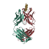
| ||||||||
| 2 | 
| ||||||||
| Unit cell |
|
-Components
| #1: Antibody | Mass: 23300.748 Da / Num. of mol.: 2 Source method: isolated from a genetically manipulated source Source: (gene. exp.) Homo sapiens (human) / Cell line (production host): Expi 293F / Production host: Homo sapiens (human)#2: Antibody | Mass: 23913.920 Da / Num. of mol.: 2 Source method: isolated from a genetically manipulated source Source: (gene. exp.) Homo sapiens (human) / Cell line (production host): Expi 293F / Production host: Homo sapiens (human)#3: Protein/peptide | Mass: 2187.582 Da / Num. of mol.: 2 / Source method: obtained synthetically / Source: (synth.) Human immunodeficiency virus 1 / References: UniProt: P04578*PLUS#4: Chemical |   Mass: 368.360 Da / Num. of mol.: 2 / Source method: obtained synthetically / Formula: C15H29O8P / Comment: phospholipid*YM Mass: 368.360 Da / Num. of mol.: 2 / Source method: obtained synthetically / Formula: C15H29O8P / Comment: phospholipid*YM#5: Chemical | ChemComp-MPD / ( | 2-Methyl-2,4-pentanediol  Mass: 118.174 Da / Num. of mol.: 1 / Source method: obtained synthetically / Formula: C6H14O2 / Comment: precipitant*YM Mass: 118.174 Da / Num. of mol.: 1 / Source method: obtained synthetically / Formula: C6H14O2 / Comment: precipitant*YM |
|---|
-Experimental details
-Experiment
| Experiment | Method: X-RAY DIFFRACTION / Number of used crystals: 1 |
|---|
- Sample preparation
| Crystal | Density Matthews: 3.17 Å3/Da / Density % sol: 60.9 % |
|---|---|
| Crystal grow | Temperature: 293.15 K / Method: vapor diffusion, sitting drop / pH: 5.5 Details: Reservoir: 0.1 M sodium acetate pH 5.5, 12% 2-methyl-2,4-pentanediol |
-Data collection
| Diffraction | Mean temperature: 100 K / Serial crystal experiment: N |
|---|---|
| Diffraction source | Source: SYNCHROTRON / Site: SSRL / Beamline: BL9-2 / Wavelength: 0.97946 Å |
| Detector | Type: DECTRIS PILATUS 6M / Detector: PIXEL / Date: Apr 18, 2017 |
| Radiation | Protocol: SINGLE WAVELENGTH / Monochromatic (M) / Laue (L): M / Scattering type: x-ray |
| Radiation wavelength | Wavelength: 0.97946 Å / Relative weight: 1 |
| Reflection | Resolution: 3.416→44.1 Å / Num. obs: 15913 / % possible obs: 93.4 % / Redundancy: 4.4 % / Biso Wilson estimate: 79 Å2 / CC1/2: 0.875 / Rpim(I) all: 0.11 / Rsym value: 0.21 / Net I/σ(I): 6.6 |
| Reflection shell | Resolution: 3.416→3.48 Å / Redundancy: 4.4 % / Mean I/σ(I) obs: 1.8 / Num. unique obs: 810 / CC1/2: 0.572 / Rpim(I) all: 0.466 / Rsym value: 0.887 / % possible all: 95.3 |
- Processing
| Software |
| |||||||||||||||||||||||||||||||||||||||||||||||||
|---|---|---|---|---|---|---|---|---|---|---|---|---|---|---|---|---|---|---|---|---|---|---|---|---|---|---|---|---|---|---|---|---|---|---|---|---|---|---|---|---|---|---|---|---|---|---|---|---|---|---|
| Refinement | Method to determine structure: MOLECULAR REPLACEMENT Starting model: 6O3D Resolution: 3.416→44.097 Å / SU ML: 0.43 / Cross valid method: FREE R-VALUE / σ(F): 1.34 / Phase error: 27.16
| |||||||||||||||||||||||||||||||||||||||||||||||||
| Solvent computation | Shrinkage radii: 0.9 Å / VDW probe radii: 1.11 Å | |||||||||||||||||||||||||||||||||||||||||||||||||
| Displacement parameters | Biso mean: 79 Å2 | |||||||||||||||||||||||||||||||||||||||||||||||||
| Refinement step | Cycle: LAST / Resolution: 3.416→44.097 Å
| |||||||||||||||||||||||||||||||||||||||||||||||||
| Refine LS restraints |
| |||||||||||||||||||||||||||||||||||||||||||||||||
| LS refinement shell |
|
