 Movie
Movie Controller

[English] 日本語
 Yorodumi
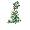
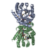
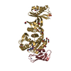
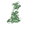
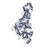
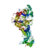
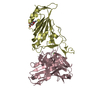
- PDB-6o1x: Structure of pCW3 conjugation coupling protein TcpA monomer form ... -
+ Open data

- Basic information
| Entry | Database: PDB / ID: 6o1x | |||||||||
|---|---|---|---|---|---|---|---|---|---|---|
| Title | Structure of pCW3 conjugation coupling protein TcpA monomer form with ATPgS | |||||||||
 Components Components | DNA translocase coupling protein | |||||||||
 Keywords Keywords |  TRANSLOCASE / ATPase / Conjugation TRANSLOCASE / ATPase / Conjugation | |||||||||
| Function / homology |  Function and homology information Function and homology information | |||||||||
| Biological species |   Clostridium perfringens (bacteria) Clostridium perfringens (bacteria) | |||||||||
| Method | X-RAY DIFFRACTION / SYNCHROTRON / MOLECULAR REPLACEMENT / molecular replacement / Resolution: 2.46 Å | |||||||||
 Authors Authors | Traore, D.A.K. / Ahktar, N. / Torres, V.T. / Adams, V. / Coulibaly, F. / Panjikar, S. / Caradoc-Davies, T.T. / Rood, J.I. / Whisstock, J.C. | |||||||||
| Funding support |  Australia, 1items Australia, 1items
| |||||||||
 Citation Citation | Journal: To be published Title: Structure of pCW3 conjugation coupling protein TcpA Authors: Traore, D.A.K. / Whisstock, J.C. | |||||||||
| History |
|
- Structure visualization
| Structure viewer | Molecule: MolmilJmol/JSmol |
|---|
- Downloads & links
-Download
| PDBx/mmCIF format | 6o1x.cif.gz | 296.5 KB | Display | PDBx/mmCIF format |
|---|---|---|---|---|
| PDB format | pdb6o1x.ent.gz | 238.2 KB | Display | PDB format |
| PDBx/mmJSON format | 6o1x.json.gz | Tree view | PDBx/mmJSON format | |
| Others |  Other downloads Other downloads |
-Validation report
| Arichive directory | https://data.pdbj.org/pub/pdb/validation_reports/o1/6o1xftp://data.pdbj.org/pub/pdb/validation_reports/o1/6o1x | HTTPS FTP |
|---|
-Related structure data
| Related structure data | 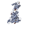 6o1wSC 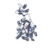 6o1yC 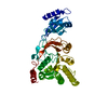 6o1zC S: Starting model for refinement C: citing same article ( |
|---|---|
| Similar structure data |


-Links
 PDBj
PDBj


- Assembly
| Deposited unit | 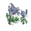
| ||||||||
|---|---|---|---|---|---|---|---|---|---|
| 1 | 
| ||||||||
| 2 | 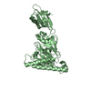
| ||||||||
| Unit cell |
|
-Components
| #1: Protein | Mass: 40435.590 Da / Num. of mol.: 2 Source method: isolated from a genetically manipulated source Source: (gene. exp.) Clostridium perfringens (bacteria) / Gene: tcpA, pCW3_0030 / Production host: Escherichia coli (E. coli) / References: UniProt: Q1PLI0#2: Sugar | Glucose  Type: D-saccharide, beta linking / Mass: 180.156 Da / Num. of mol.: 2 Type: D-saccharide, beta linking / Mass: 180.156 Da / Num. of mol.: 2Source method: isolated from a genetically manipulated source Formula: C6H12O6 #3: Chemical | ChemComp-AGS / |   Mass: 523.247 Da / Num. of mol.: 1 / Source method: obtained synthetically / Formula: C10H16N5O12P3S / Comment: ATP-gamma-S, energy-carrying molecule analogue*YM Mass: 523.247 Da / Num. of mol.: 1 / Source method: obtained synthetically / Formula: C10H16N5O12P3S / Comment: ATP-gamma-S, energy-carrying molecule analogue*YM#4: Water | ChemComp-HOH / | Water Mass: 18.015 Da / Num. of mol.: 184 / Source method: isolated from a natural source / Formula: H2O Mass: 18.015 Da / Num. of mol.: 184 / Source method: isolated from a natural source / Formula: H2O |
|---|
-Experimental details
-Experiment
| Experiment | Method: X-RAY DIFFRACTION / Number of used crystals: 1 |
|---|
- Sample preparation
| Crystal | Density Matthews: 2.14 Å3/Da / Density % sol: 42.51 % |
|---|---|
| Crystal grow | Temperature: 293 K / Method: vapor diffusion, hanging drop / pH: 5.5 Details: 20 % (w/v) PEG 3350, 0.1 M Bis-Tris pH 5.5, 0.2 M NaCl |
-Data collection
| Diffraction | Mean temperature: 100 K / Serial crystal experiment: N | ||||||||||||||||||||||||||||||
|---|---|---|---|---|---|---|---|---|---|---|---|---|---|---|---|---|---|---|---|---|---|---|---|---|---|---|---|---|---|---|---|
| Diffraction source | Source: SYNCHROTRON / Site: Australian Synchrotron / Beamline: MX2 / Wavelength: 0.9737 Å | ||||||||||||||||||||||||||||||
| Detector | Type: DECTRIS EIGER X 16M / Detector: PIXEL / Date: Apr 24, 2018 | ||||||||||||||||||||||||||||||
| Radiation | Protocol: SINGLE WAVELENGTH / Monochromatic (M) / Laue (L): M / Scattering type: x-ray | ||||||||||||||||||||||||||||||
| Radiation wavelength | Wavelength: 0.9737 Å / Relative weight: 1 | ||||||||||||||||||||||||||||||
| Reflection | Resolution: 2.46→48.97 Å / Num. obs: 25446 / % possible obs: 99.8 % / Redundancy: 9 % / Biso Wilson estimate: 52.09 Å2 / CC1/2: 0.998 / Rmerge(I) obs: 0.109 / Rpim(I) all: 0.038 / Rrim(I) all: 0.115 / Net I/σ(I): 13 / Num. measured all: 228939 / Scaling rejects: 23 | ||||||||||||||||||||||||||||||
| Reflection shell | Diffraction-ID: 1
|
-Phasing
| Phasing | Method: molecular replacement |
|---|
- Processing
| Software |
| ||||||||||||||||||||||||||||||||||||||||||||||||||||||||||||||||||||||||||||||||||||||||||||||||||||||||||||
|---|---|---|---|---|---|---|---|---|---|---|---|---|---|---|---|---|---|---|---|---|---|---|---|---|---|---|---|---|---|---|---|---|---|---|---|---|---|---|---|---|---|---|---|---|---|---|---|---|---|---|---|---|---|---|---|---|---|---|---|---|---|---|---|---|---|---|---|---|---|---|---|---|---|---|---|---|---|---|---|---|---|---|---|---|---|---|---|---|---|---|---|---|---|---|---|---|---|---|---|---|---|---|---|---|---|---|---|---|---|
| Refinement | Method to determine structure: MOLECULAR REPLACEMENT Starting model: 6O1W Resolution: 2.46→46.2 Å / Cor.coef. Fo:Fc: 0.947 / Cor.coef. Fo:Fc free: 0.915 / SU R Cruickshank DPI: 0.696 / Cross valid method: THROUGHOUT / σ(F): 0 / SU R Blow DPI: 0.835 / SU Rfree Blow DPI: 0.274 / SU Rfree Cruickshank DPI: 0.275
| ||||||||||||||||||||||||||||||||||||||||||||||||||||||||||||||||||||||||||||||||||||||||||||||||||||||||||||
| Displacement parameters | Biso max: 134.42 Å2 / Biso mean: 55.59 Å2 / Biso min: 15.7 Å2
| ||||||||||||||||||||||||||||||||||||||||||||||||||||||||||||||||||||||||||||||||||||||||||||||||||||||||||||
| Refine analyze | Luzzati coordinate error obs: 0.27 Å | ||||||||||||||||||||||||||||||||||||||||||||||||||||||||||||||||||||||||||||||||||||||||||||||||||||||||||||
| Refinement step | Cycle: final / Resolution: 2.46→46.2 Å
| ||||||||||||||||||||||||||||||||||||||||||||||||||||||||||||||||||||||||||||||||||||||||||||||||||||||||||||
| Refine LS restraints |
| ||||||||||||||||||||||||||||||||||||||||||||||||||||||||||||||||||||||||||||||||||||||||||||||||||||||||||||
| LS refinement shell | Resolution: 2.46→2.48 Å / Rfactor Rfree error: 0 / Total num. of bins used: 50
| ||||||||||||||||||||||||||||||||||||||||||||||||||||||||||||||||||||||||||||||||||||||||||||||||||||||||||||
| Refinement TLS params. | Method: refined / Refine-ID: X-RAY DIFFRACTION
| ||||||||||||||||||||||||||||||||||||||||||||||||||||||||||||||||||||||||||||||||||||||||||||||||||||||||||||
| Refinement TLS group |
|
