 Movie
Movie Controller

[English] 日本語
 Yorodumi
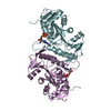
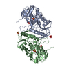
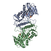
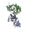
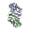
- PDB-6nfe: Crystal Structure of Ribose-phosphate Pyrophosphokinase from Legi... -
+ Open data

- Basic information
| Entry | Database: PDB / ID: 6nfe | |||||||||
|---|---|---|---|---|---|---|---|---|---|---|
| Title | Crystal Structure of Ribose-phosphate Pyrophosphokinase from Legionella pneumophila with bound AMP, ADP, and Ribose-5-Phosphate | |||||||||
 Components Components | Ribose-phosphate pyrophosphokinase | |||||||||
 Keywords Keywords |  LIGASE / SSGCID / phosphorylation / AMP / ADP / Ribose-5-Phosphate / Structural Genomics / Seattle Structural Genomics Center for Infectious Disease LIGASE / SSGCID / phosphorylation / AMP / ADP / Ribose-5-Phosphate / Structural Genomics / Seattle Structural Genomics Center for Infectious Disease | |||||||||
| Function / homology |  Function and homology informationribose-phosphate diphosphokinase / ribose phosphate diphosphokinase activity / ribonucleoside monophosphate biosynthetic process / nucleotide biosynthetic process / 5-phosphoribose 1-diphosphate biosynthetic process / nucleoside metabolic process / kinase activity / magnesium ion binding / ATP binding / cytoplasm Function and homology informationribose-phosphate diphosphokinase / ribose phosphate diphosphokinase activity / ribonucleoside monophosphate biosynthetic process / nucleotide biosynthetic process / 5-phosphoribose 1-diphosphate biosynthetic process / nucleoside metabolic process / kinase activity / magnesium ion binding / ATP binding / cytoplasmSimilarity search - Function | |||||||||
| Biological species |   Legionella pneumophila (bacteria) Legionella pneumophila (bacteria) | |||||||||
| Method | X-RAY DIFFRACTION / SYNCHROTRON / MOLECULAR REPLACEMENT / molecular replacement / Resolution: 1.75 Å | |||||||||
 Authors Authors | Seattle Structural Genomics Center for Infectious Disease (SSGCID) | |||||||||
 Citation Citation | Journal: to be published Title: Crystal Structure of Ribose-phosphate Pyrophosphokinase from Legionella pneumophila with bound AMP, ADP, and Ribose-5-Phosphate Authors: Bolejack, M.J. / Abendroth, J. / Lorimer, D.D. / Horanyi, P.S. / Edwards, T.E. | |||||||||
| History |
|
- Structure visualization
| Structure viewer | Molecule: MolmilJmol/JSmol |
|---|
- Downloads & links
-Download
| PDBx/mmCIF format | 6nfe.cif.gz | 259.9 KB | Display | PDBx/mmCIF format |
|---|---|---|---|---|
| PDB format | pdb6nfe.ent.gz | 207.1 KB | Display | PDB format |
| PDBx/mmJSON format | 6nfe.json.gz | Tree view | PDBx/mmJSON format | |
| Others |  Other downloads Other downloads |
-Validation report
| Arichive directory | https://data.pdbj.org/pub/pdb/validation_reports/nf/6nfeftp://data.pdbj.org/pub/pdb/validation_reports/nf/6nfe | HTTPS FTP |
|---|
-Related structure data
| Related structure data | 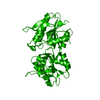 4s2uS S: Starting model for refinement |
|---|---|
| Similar structure data | |
| Other databases |


-Links
 PDBj
PDBj


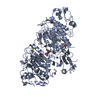
- Assembly
| Deposited unit | 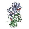
| ||||||||
|---|---|---|---|---|---|---|---|---|---|
| 1 |
| ||||||||
| Unit cell |
|
-Components
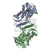
-Protein / Sugars , 2 types, 4 molecules AB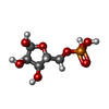
| #1: Protein | Mass: 35199.676 Da / Num. of mol.: 2 / Fragment: LepnA.00035.a.B1 Source method: isolated from a genetically manipulated source Source: (gene. exp.) Legionella pneumophila (bacteria) / Gene: prsA, prs, C3927_02005, NCTC12024_00599 / Plasmid: LepnA.00035.a.B1 / Production host: Escherichia coli (E. coli) / Strain (production host): BL21(DE3)References: UniProt: A0A2S6FBX1, UniProt: Q5ZY30*PLUS, ribose-phosphate diphosphokinase#4: Sugar |  Type: D-saccharide, alpha linking / Mass: 230.110 Da / Num. of mol.: 2 Type: D-saccharide, alpha linking / Mass: 230.110 Da / Num. of mol.: 2Source method: isolated from a genetically manipulated source Formula: C5H11O8P |
|---|
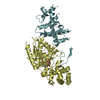
-Non-polymers , 5 types, 542 molecules 


| #2: Chemical | ChemComp-EDO / Ethylene glycol Mass: 62.068 Da / Num. of mol.: 11 / Source method: obtained synthetically / Formula: C2H6O2 Mass: 62.068 Da / Num. of mol.: 11 / Source method: obtained synthetically / Formula: C2H6O2#3: Chemical | Ethanol Mass: 46.068 Da / Num. of mol.: 3 / Source method: obtained synthetically / Formula: C2H6O Mass: 46.068 Da / Num. of mol.: 3 / Source method: obtained synthetically / Formula: C2H6O#5: Chemical | Adenosine monophosphate Mass: 347.221 Da / Num. of mol.: 2 / Source method: obtained synthetically / Formula: C10H14N5O7P / Comment: AMP*YM Mass: 347.221 Da / Num. of mol.: 2 / Source method: obtained synthetically / Formula: C10H14N5O7P / Comment: AMP*YM#6: Chemical | Adenosine diphosphate Mass: 427.201 Da / Num. of mol.: 2 / Source method: obtained synthetically / Formula: C10H15N5O10P2 / Comment: ADP, energy-carrying molecule*YM Mass: 427.201 Da / Num. of mol.: 2 / Source method: obtained synthetically / Formula: C10H15N5O10P2 / Comment: ADP, energy-carrying molecule*YM#7: Water | ChemComp-HOH / | WaterMass: 18.015 Da / Num. of mol.: 524 / Source method: isolated from a natural source / Formula: H2O |
|---|
-Experimental details
-Experiment
| Experiment | Method: X-RAY DIFFRACTION / Number of used crystals: 1 |
|---|
- Sample preparation
| Crystal | Density Matthews: 2.37 Å3/Da / Density % sol: 48.1 % |
|---|---|
| Crystal grow | Temperature: 290 K / Method: vapor diffusion, sitting drop / pH: 4.2 Details: LepnA.00035.a.B1.PS38405 at 19.13 mg/ml was incubated with 4 mM ribose-5-phosphate, 4 mM ATP, and 4 mM MgCl2 then was mixed 1:1 JCSG+(b6): 40% (v/v) reagent alcohol, 100 mM sodium phosphate ...Details: LepnA.00035.a.B1.PS38405 at 19.13 mg/ml was incubated with 4 mM ribose-5-phosphate, 4 mM ATP, and 4 mM MgCl2 then was mixed 1:1 JCSG+(b6): 40% (v/v) reagent alcohol, 100 mM sodium phosphate dibasic/citric acid pH 4.2. Tray: 298764b6, puck: uya9-9. |
-Data collection
| Diffraction | Mean temperature: 100 K / Serial crystal experiment: N | ||||||||||||||||||||||||||||||||||||||||||||||||||||||||||||||||||||||||||||||||||||||||||||||||||||||||||||||||||||||||||||||||||||||||||||||||||||||||||||||||||||||||||||||||||||||||||||||||||||||||||||||||||
|---|---|---|---|---|---|---|---|---|---|---|---|---|---|---|---|---|---|---|---|---|---|---|---|---|---|---|---|---|---|---|---|---|---|---|---|---|---|---|---|---|---|---|---|---|---|---|---|---|---|---|---|---|---|---|---|---|---|---|---|---|---|---|---|---|---|---|---|---|---|---|---|---|---|---|---|---|---|---|---|---|---|---|---|---|---|---|---|---|---|---|---|---|---|---|---|---|---|---|---|---|---|---|---|---|---|---|---|---|---|---|---|---|---|---|---|---|---|---|---|---|---|---|---|---|---|---|---|---|---|---|---|---|---|---|---|---|---|---|---|---|---|---|---|---|---|---|---|---|---|---|---|---|---|---|---|---|---|---|---|---|---|---|---|---|---|---|---|---|---|---|---|---|---|---|---|---|---|---|---|---|---|---|---|---|---|---|---|---|---|---|---|---|---|---|---|---|---|---|---|---|---|---|---|---|---|---|---|---|---|---|---|
| Diffraction source | Source: SYNCHROTRON / Site: APS  / Beamline: 21-ID-F / Wavelength: 0.97872 Å / Beamline: 21-ID-F / Wavelength: 0.97872 Å | ||||||||||||||||||||||||||||||||||||||||||||||||||||||||||||||||||||||||||||||||||||||||||||||||||||||||||||||||||||||||||||||||||||||||||||||||||||||||||||||||||||||||||||||||||||||||||||||||||||||||||||||||||
| Detector | Type: RAYONIX MX-300 / Detector: CCD / Date: Apr 12, 2018 / Details: Beryllium Lenses | ||||||||||||||||||||||||||||||||||||||||||||||||||||||||||||||||||||||||||||||||||||||||||||||||||||||||||||||||||||||||||||||||||||||||||||||||||||||||||||||||||||||||||||||||||||||||||||||||||||||||||||||||||
| Radiation | Monochromator: Diamond [111] / Protocol: SINGLE WAVELENGTH / Monochromatic (M) / Laue (L): M / Scattering type: x-ray | ||||||||||||||||||||||||||||||||||||||||||||||||||||||||||||||||||||||||||||||||||||||||||||||||||||||||||||||||||||||||||||||||||||||||||||||||||||||||||||||||||||||||||||||||||||||||||||||||||||||||||||||||||
| Radiation wavelength | Wavelength: 0.97872 Å / Relative weight: 1 | ||||||||||||||||||||||||||||||||||||||||||||||||||||||||||||||||||||||||||||||||||||||||||||||||||||||||||||||||||||||||||||||||||||||||||||||||||||||||||||||||||||||||||||||||||||||||||||||||||||||||||||||||||
| Reflection | Resolution: 1.75→43.245 Å / Num. obs: 67559 / % possible obs: 99.9 % / Redundancy: 7.462 % / Biso Wilson estimate: 26.218 Å2 / CC1/2: 0.998 / Rmerge(I) obs: 0.072 / Rrim(I) all: 0.077 / Χ2: 1.02 / Net I/σ(I): 18.78 / Num. measured all: 504098 | ||||||||||||||||||||||||||||||||||||||||||||||||||||||||||||||||||||||||||||||||||||||||||||||||||||||||||||||||||||||||||||||||||||||||||||||||||||||||||||||||||||||||||||||||||||||||||||||||||||||||||||||||||
| Reflection shell | Diffraction-ID: 1
|
-Phasing
| Phasing | Method: molecular replacement |
|---|
- Processing
| Software |
| |||||||||||||||||||||||||||||||||||||||||||||||||||||||||||||||||||||||||||||||||||||||||||||||||||||||||||||||||||||||||||||||||||||||||||||||||||||||||||||||||||||||||||||||||||||||||||||||||||||||||||||||||||||||||||||||||||||||||||||||||||||||||||||||||||||||||||||||||||||||||||||||||||||||||||||||||||||||||||||||||||||||||||||||||||||||||||||||||||||||||||||||||||||||||||||||||||||||||||||||||||||||||||||||||||||||||
|---|---|---|---|---|---|---|---|---|---|---|---|---|---|---|---|---|---|---|---|---|---|---|---|---|---|---|---|---|---|---|---|---|---|---|---|---|---|---|---|---|---|---|---|---|---|---|---|---|---|---|---|---|---|---|---|---|---|---|---|---|---|---|---|---|---|---|---|---|---|---|---|---|---|---|---|---|---|---|---|---|---|---|---|---|---|---|---|---|---|---|---|---|---|---|---|---|---|---|---|---|---|---|---|---|---|---|---|---|---|---|---|---|---|---|---|---|---|---|---|---|---|---|---|---|---|---|---|---|---|---|---|---|---|---|---|---|---|---|---|---|---|---|---|---|---|---|---|---|---|---|---|---|---|---|---|---|---|---|---|---|---|---|---|---|---|---|---|---|---|---|---|---|---|---|---|---|---|---|---|---|---|---|---|---|---|---|---|---|---|---|---|---|---|---|---|---|---|---|---|---|---|---|---|---|---|---|---|---|---|---|---|---|---|---|---|---|---|---|---|---|---|---|---|---|---|---|---|---|---|---|---|---|---|---|---|---|---|---|---|---|---|---|---|---|---|---|---|---|---|---|---|---|---|---|---|---|---|---|---|---|---|---|---|---|---|---|---|---|---|---|---|---|---|---|---|---|---|---|---|---|---|---|---|---|---|---|---|---|---|---|---|---|---|---|---|---|---|---|---|---|---|---|---|---|---|---|---|---|---|---|---|---|---|---|---|---|---|---|---|---|---|---|---|---|---|---|---|---|---|---|---|---|---|---|---|---|---|---|---|---|---|---|---|---|---|---|---|---|---|---|---|---|---|---|---|---|---|---|---|---|---|---|---|---|---|---|---|---|---|---|---|---|---|---|---|---|---|---|---|---|---|---|---|---|---|---|---|---|---|---|---|---|---|---|---|---|---|---|---|---|---|---|---|---|---|---|---|---|---|---|---|---|---|---|---|---|---|---|---|---|---|---|---|---|---|---|
| Refinement | Method to determine structure: MOLECULAR REPLACEMENT Starting model: 4S2U Resolution: 1.75→43.245 Å / SU ML: 0.17 / Cross valid method: FREE R-VALUE / σ(F): 1.36 / Phase error: 16.37
| |||||||||||||||||||||||||||||||||||||||||||||||||||||||||||||||||||||||||||||||||||||||||||||||||||||||||||||||||||||||||||||||||||||||||||||||||||||||||||||||||||||||||||||||||||||||||||||||||||||||||||||||||||||||||||||||||||||||||||||||||||||||||||||||||||||||||||||||||||||||||||||||||||||||||||||||||||||||||||||||||||||||||||||||||||||||||||||||||||||||||||||||||||||||||||||||||||||||||||||||||||||||||||||||||||||||||
| Solvent computation | Shrinkage radii: 0.9 Å / VDW probe radii: 1.11 Å | |||||||||||||||||||||||||||||||||||||||||||||||||||||||||||||||||||||||||||||||||||||||||||||||||||||||||||||||||||||||||||||||||||||||||||||||||||||||||||||||||||||||||||||||||||||||||||||||||||||||||||||||||||||||||||||||||||||||||||||||||||||||||||||||||||||||||||||||||||||||||||||||||||||||||||||||||||||||||||||||||||||||||||||||||||||||||||||||||||||||||||||||||||||||||||||||||||||||||||||||||||||||||||||||||||||||||
| Displacement parameters | Biso max: 101.36 Å2 / Biso mean: 25.2569 Å2 / Biso min: 8.04 Å2 | |||||||||||||||||||||||||||||||||||||||||||||||||||||||||||||||||||||||||||||||||||||||||||||||||||||||||||||||||||||||||||||||||||||||||||||||||||||||||||||||||||||||||||||||||||||||||||||||||||||||||||||||||||||||||||||||||||||||||||||||||||||||||||||||||||||||||||||||||||||||||||||||||||||||||||||||||||||||||||||||||||||||||||||||||||||||||||||||||||||||||||||||||||||||||||||||||||||||||||||||||||||||||||||||||||||||||
| Refinement step | Cycle: final / Resolution: 1.75→43.245 Å
| |||||||||||||||||||||||||||||||||||||||||||||||||||||||||||||||||||||||||||||||||||||||||||||||||||||||||||||||||||||||||||||||||||||||||||||||||||||||||||||||||||||||||||||||||||||||||||||||||||||||||||||||||||||||||||||||||||||||||||||||||||||||||||||||||||||||||||||||||||||||||||||||||||||||||||||||||||||||||||||||||||||||||||||||||||||||||||||||||||||||||||||||||||||||||||||||||||||||||||||||||||||||||||||||||||||||||
| LS refinement shell | Refine-ID: X-RAY DIFFRACTION / Rfactor Rfree error: 0 / Total num. of bins used: 14
| |||||||||||||||||||||||||||||||||||||||||||||||||||||||||||||||||||||||||||||||||||||||||||||||||||||||||||||||||||||||||||||||||||||||||||||||||||||||||||||||||||||||||||||||||||||||||||||||||||||||||||||||||||||||||||||||||||||||||||||||||||||||||||||||||||||||||||||||||||||||||||||||||||||||||||||||||||||||||||||||||||||||||||||||||||||||||||||||||||||||||||||||||||||||||||||||||||||||||||||||||||||||||||||||||||||||||
| Refinement TLS params. | Method: refined / Refine-ID: X-RAY DIFFRACTION
| |||||||||||||||||||||||||||||||||||||||||||||||||||||||||||||||||||||||||||||||||||||||||||||||||||||||||||||||||||||||||||||||||||||||||||||||||||||||||||||||||||||||||||||||||||||||||||||||||||||||||||||||||||||||||||||||||||||||||||||||||||||||||||||||||||||||||||||||||||||||||||||||||||||||||||||||||||||||||||||||||||||||||||||||||||||||||||||||||||||||||||||||||||||||||||||||||||||||||||||||||||||||||||||||||||||||||
| Refinement TLS group |
|