 Movie
Movie Controller

[English] 日本語
 Yorodumi
- PDB-3mbi: Crystal structure of the phosphoribosylpyrophosphate (PRPP) synth... -
+ Open data

- Basic information
| Entry | Database: PDB / ID: 3mbi | |||||||||
|---|---|---|---|---|---|---|---|---|---|---|
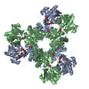
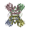
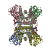
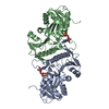
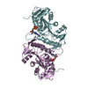
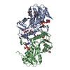
| Title | Crystal structure of the phosphoribosylpyrophosphate (PRPP) synthetase from Thermoplasma volcanium in complex with ADP-Mg2+ and ribose 5-phosphate | |||||||||
 Components Components | Ribose-phosphate pyrophosphokinase | |||||||||
 Keywords Keywords |  TRANSFERASE / Phosphoribosyl transferase / ATP analog binding / Ribose 5-phosphate binding TRANSFERASE / Phosphoribosyl transferase / ATP analog binding / Ribose 5-phosphate binding | |||||||||
| Function / homology |  Function and homology informationribose-phosphate diphosphokinase / ribose phosphate diphosphokinase activity / ribonucleoside monophosphate biosynthetic process / nucleotide biosynthetic process / 5-phosphoribose 1-diphosphate biosynthetic process / kinase activity / phosphorylation / magnesium ion binding / ATP binding / cytoplasm Function and homology informationribose-phosphate diphosphokinase / ribose phosphate diphosphokinase activity / ribonucleoside monophosphate biosynthetic process / nucleotide biosynthetic process / 5-phosphoribose 1-diphosphate biosynthetic process / kinase activity / phosphorylation / magnesium ion binding / ATP binding / cytoplasmSimilarity search - Function | |||||||||
| Biological species |   Thermoplasma volcanium (archaea) Thermoplasma volcanium (archaea) | |||||||||
| Method | X-RAY DIFFRACTION / SYNCHROTRON / MOLECULAR REPLACEMENT / Resolution: 1.8 Å | |||||||||
 Authors Authors | Cherney, L.T. / Cherney, M.M. / Garen, C.R. / James, M.N.G. | |||||||||
 Citation Citation | Journal: J.Mol.Biol. / Year: 2011 Title: The structures of Thermoplasma volcanium phosphoribosyl pyrophosphate synthetase bound to ribose-5-phosphate and ATP analogs. Authors: Cherney, M.M. / Cherney, L.T. / Garen, C.R. / James, M.N. | |||||||||
| History |
|
- Structure visualization
| Structure viewer | Molecule: MolmilJmol/JSmol |
|---|
- Downloads & links
-Download
| PDBx/mmCIF format | 3mbi.cif.gz | 265.2 KB | Display | PDBx/mmCIF format |
|---|---|---|---|---|
| PDB format | pdb3mbi.ent.gz | 211.6 KB | Display | PDB format |
| PDBx/mmJSON format | 3mbi.json.gz | Tree view | PDBx/mmJSON format | |
| Others |  Other downloads Other downloads |
-Validation report
| Arichive directory | https://data.pdbj.org/pub/pdb/validation_reports/mb/3mbiftp://data.pdbj.org/pub/pdb/validation_reports/mb/3mbi | HTTPS FTP |
|---|
-Related structure data
| Related structure data |  3lpnSC 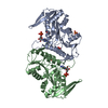 3lrtC 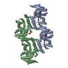 3nagC C: citing same article ( S: Starting model for refinement |
|---|---|
| Similar structure data |


-Links
 PDBj
PDBj
- Assembly
| Deposited unit | 
| ||||||||
|---|---|---|---|---|---|---|---|---|---|
| 1 | 
| ||||||||
| 2 | 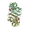
| ||||||||
| Unit cell |
|
-Components
-Protein / Sugars , 2 types, 5 molecules ABCD
| #1: Protein | Mass: 32294.492 Da / Num. of mol.: 4 Source method: isolated from a genetically manipulated source Source: (gene. exp.) Thermoplasma volcanium (archaea) / Strain: ATCC51530 / Gene: prs, TV0197, TVG0201915, TVN0197 / Plasmid: pvp16 / Production host:  Escherichia coli (E. coli) / Strain (production host): BL21 (DE3) PlysS Escherichia coli (E. coli) / Strain (production host): BL21 (DE3) PlysSReferences: UniProt: Q97CA5, ribose-phosphate diphosphokinase#5: Sugar | ChemComp-HSX / |  Type: D-saccharide, alpha linking / Mass: 230.110 Da / Num. of mol.: 1 Type: D-saccharide, alpha linking / Mass: 230.110 Da / Num. of mol.: 1Source method: isolated from a genetically manipulated source Formula: C5H11O8P |
|---|
-Non-polymers , 4 types, 1265 molecules 


| #2: Chemical | Phosphate Mass: 94.971 Da / Num. of mol.: 3 / Source method: obtained synthetically / Formula: PO4 Mass: 94.971 Da / Num. of mol.: 3 / Source method: obtained synthetically / Formula: PO4#3: Chemical | ChemComp-MG /  Mass: 24.305 Da / Num. of mol.: 4 / Source method: obtained synthetically / Formula: Mg Mass: 24.305 Da / Num. of mol.: 4 / Source method: obtained synthetically / Formula: Mg#4: Chemical | ChemComp-ADP / Adenosine diphosphate Mass: 427.201 Da / Num. of mol.: 4 / Source method: obtained synthetically / Formula: C10H15N5O10P2 / Comment: ADP, energy-carrying molecule*YM Mass: 427.201 Da / Num. of mol.: 4 / Source method: obtained synthetically / Formula: C10H15N5O10P2 / Comment: ADP, energy-carrying molecule*YM#6: Water | ChemComp-HOH / | WaterMass: 18.015 Da / Num. of mol.: 1254 / Source method: isolated from a natural source / Formula: H2O |
|---|
-Experimental details
-Experiment
| Experiment | Method: X-RAY DIFFRACTION / Number of used crystals: 1 |
|---|
- Sample preparation
| Crystal | Density Matthews: 2.28 Å3/Da / Density % sol: 45.97 % |
|---|---|
| Crystal grow | Temperature: 293 K / pH: 7.5 Details: 30% PEG600, 0.1M HEPES, 10% MPD, 0.1% LDAO, pH 7.5, VAPOR DIFFUSION, HANGING DROP, temperature 293K |
-Data collection
| Diffraction | Mean temperature: 100 K |
|---|---|
| Diffraction source | Source: SYNCHROTRON / Site: CLSI  / Beamline: 08ID-1 / Wavelength: 0.9795 / Beamline: 08ID-1 / Wavelength: 0.9795 |
| Detector | Type: MARMOSAIC 225 mm CCD / Detector: CCD / Date: Aug 25, 2009 |
| Radiation | Protocol: SINGLE WAVELENGTH / Monochromatic (M) / Laue (L): M / Scattering type: x-ray |
| Radiation wavelength | Wavelength: 0.9795 Å / Relative weight: 1 |
| Reflection | Resolution: 1.797→25.471 Å / Num. obs: 105923 / % possible obs: 97.8 % / Observed criterion σ(I): 0 / Redundancy: 3.9 % / Biso Wilson estimate: 16.2 Å2 / Rmerge(I) obs: 0.12 / Rsym value: 0.12 / Net I/σ(I): 11.8 |
| Reflection shell | Resolution: 1.8→1.86 Å / Redundancy: 3.8 % / Rmerge(I) obs: 0.71 / Mean I/σ(I) obs: 1.8 / Rsym value: 0.71 / % possible all: 96.4 |
- Processing
| Software |
| |||||||||||||||||||||||||||||||||||||||||||||||||||||||||||||||||||||||||||||||||||||||||||||||||||||||||||||||||||||||||||||||||||||||||||||||||||||||||||||||||||||||||||||||||||||||||||||||||||||||||||||||||||||||||
|---|---|---|---|---|---|---|---|---|---|---|---|---|---|---|---|---|---|---|---|---|---|---|---|---|---|---|---|---|---|---|---|---|---|---|---|---|---|---|---|---|---|---|---|---|---|---|---|---|---|---|---|---|---|---|---|---|---|---|---|---|---|---|---|---|---|---|---|---|---|---|---|---|---|---|---|---|---|---|---|---|---|---|---|---|---|---|---|---|---|---|---|---|---|---|---|---|---|---|---|---|---|---|---|---|---|---|---|---|---|---|---|---|---|---|---|---|---|---|---|---|---|---|---|---|---|---|---|---|---|---|---|---|---|---|---|---|---|---|---|---|---|---|---|---|---|---|---|---|---|---|---|---|---|---|---|---|---|---|---|---|---|---|---|---|---|---|---|---|---|---|---|---|---|---|---|---|---|---|---|---|---|---|---|---|---|---|---|---|---|---|---|---|---|---|---|---|---|---|---|---|---|---|---|---|---|---|---|---|---|---|---|---|---|---|---|---|---|---|
| Refinement | Method to determine structure: MOLECULAR REPLACEMENT Starting model: 3LPN Resolution: 1.8→25.47 Å / SU ML: 0.22 / σ(F): 1.34 / Phase error: 22.81 / Stereochemistry target values: ML
| |||||||||||||||||||||||||||||||||||||||||||||||||||||||||||||||||||||||||||||||||||||||||||||||||||||||||||||||||||||||||||||||||||||||||||||||||||||||||||||||||||||||||||||||||||||||||||||||||||||||||||||||||||||||||
| Solvent computation | Shrinkage radii: 0.9 Å / VDW probe radii: 1.11 Å / Solvent model: FLAT BULK SOLVENT MODEL / Bsol: 39.23 Å2 / ksol: 0.33 e/Å3 | |||||||||||||||||||||||||||||||||||||||||||||||||||||||||||||||||||||||||||||||||||||||||||||||||||||||||||||||||||||||||||||||||||||||||||||||||||||||||||||||||||||||||||||||||||||||||||||||||||||||||||||||||||||||||
| Refinement step | Cycle: LAST / Resolution: 1.8→25.47 Å
| |||||||||||||||||||||||||||||||||||||||||||||||||||||||||||||||||||||||||||||||||||||||||||||||||||||||||||||||||||||||||||||||||||||||||||||||||||||||||||||||||||||||||||||||||||||||||||||||||||||||||||||||||||||||||
| Refine LS restraints |
| |||||||||||||||||||||||||||||||||||||||||||||||||||||||||||||||||||||||||||||||||||||||||||||||||||||||||||||||||||||||||||||||||||||||||||||||||||||||||||||||||||||||||||||||||||||||||||||||||||||||||||||||||||||||||
| LS refinement shell |
|
