 Movie
Movie Controller


+ Open data

- Basic information
| Entry | Database: PDB / ID: 6n64 | ||||||
|---|---|---|---|---|---|---|---|
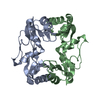
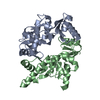
| Title | Crystal structure of mouse SMCHD1 hinge domain | ||||||
 Components Components |
| ||||||
 Keywords Keywords |  DNA BINDING PROTEIN / SMCHD1 Epigenetic control DNA binding SMC protein DNA BINDING PROTEIN / SMCHD1 Epigenetic control DNA binding SMC protein | ||||||
| Function / homology |  Function and homology information Function and homology information: / : / : / nose development / chromosome, telomeric region => GO:0000781 / Barr body / dosage compensation by inactivation of X chromosome / positive regulation of double-strand break repair via nonhomologous end joining / Hydrolases; Acting on acid anhydrides; In phosphorus-containing anhydrides / negative regulation of double-strand break repair via homologous recombination ...: / : / : / nose development / chromosome, telomeric region => GO:0000781 / Barr body / dosage compensation by inactivation of X chromosome / positive regulation of double-strand break repair via nonhomologous end joining / Hydrolases; Acting on acid anhydrides; In phosphorus-containing anhydrides / negative regulation of double-strand break repair via homologous recombination / positive regulation of DNA repair / double-strand break repair / site of double-strand break / protein homodimerization activity / ATP hydrolysis activity / DNA binding / ATP bindingSimilarity search - Function | ||||||
| Biological species |  Mus musculus (house mouse) Mus musculus (house mouse) | ||||||
| Method | X-RAY DIFFRACTION / SYNCHROTRON / SAD / Resolution: 3.3 Å | ||||||
 Authors Authors | Birkinshaw, R.W. / Chen, K. / Czabotar, P.E. / Blewitt, M.E. / Murphy, J.M. | ||||||
| Funding support |  Australia, 1items Australia, 1items
| ||||||
 Citation Citation | Journal: Sci.Signal. / Year: 2020 Title: Crystal structure of the hinge domain of Smchd1 reveals its dimerization mode and nucleic acid-binding residues. Authors: Chen, K. / Birkinshaw, R.W. / Gurzau, A.D. / Wanigasuriya, I. / Wang, R. / Iminitoff, M. / Sandow, J.J. / Young, S.N. / Hennessy, P.J. / Willson, T.A. / Heckmann, D.A. / Webb, A.I. / ...Authors: Chen, K. / Birkinshaw, R.W. / Gurzau, A.D. / Wanigasuriya, I. / Wang, R. / Iminitoff, M. / Sandow, J.J. / Young, S.N. / Hennessy, P.J. / Willson, T.A. / Heckmann, D.A. / Webb, A.I. / Blewitt, M.E. / Czabotar, P.E. / Murphy, J.M. | ||||||
| History |
|
- Structure visualization
| Structure viewer | Molecule: MolmilJmol/JSmol |
|---|
- Downloads & links
-Download
| PDBx/mmCIF format | 6n64.cif.gz | 434.5 KB | Display | PDBx/mmCIF format |
|---|---|---|---|---|
| PDB format | pdb6n64.ent.gz | 375.5 KB | Display | PDB format |
| PDBx/mmJSON format | 6n64.json.gz | Tree view | PDBx/mmJSON format | |
| Others |  Other downloads Other downloads |
-Validation report
| Arichive directory | https://data.pdbj.org/pub/pdb/validation_reports/n6/6n64ftp://data.pdbj.org/pub/pdb/validation_reports/n6/6n64 | HTTPS FTP |
|---|
-Related structure data
| Related structure data | |
|---|---|
| Similar structure data |


-Links
 PDBj
PDBj- Assembly
| Deposited unit | 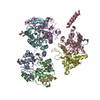
| ||||||||
|---|---|---|---|---|---|---|---|---|---|
| 1 | 
| ||||||||
| 2 | 
| ||||||||
| 3 | 
| ||||||||
| 4 | 
| ||||||||
| 5 | 
| ||||||||
| Unit cell |
|
-Components
| #1: Protein | Mass: 25388.342 Da / Num. of mol.: 6 Source method: isolated from a genetically manipulated source Source: (gene. exp.) Mus musculus (house mouse) / Gene: Smchd1, Kiaa0650 / Production host:  Escherichia coli (E. coli) Escherichia coli (E. coli)References: UniProt: Q6P5D8, Hydrolases; Acting on acid anhydrides; In phosphorus-containing anhydrides#2: Protein/peptide | Mass: 1805.216 Da / Num. of mol.: 2 Source method: isolated from a genetically manipulated source Source: (gene. exp.) Mus musculus (house mouse) / Gene: Smchd1, Kiaa0650 / Production host: Escherichia coli (E. coli)References: Hydrolases; Acting on acid anhydrides; In phosphorus-containing anhydridesHas ligand of interest | N | Sequence details | Chains G and H are portions of helix that are included in the SMCHD1 construct, but could not be ...Chains G and H are portions of helix that are included in the SMCHD1 construct, but could not be assigned to one of the chains A-F. In addition the resolution was not sufficient to dock sequence so they have been modeled as poly alanine helices, and since the registry is unknown, they are marked as UNKs. There should be 6 copies of these, but only 2 were sufficiently ordered to model as helices. | |
|---|
-Experimental details
-Experiment
| Experiment | Method: X-RAY DIFFRACTION / Number of used crystals: 1 |
|---|
- Sample preparation
| Crystal | Density Matthews: 2.85 Å3/Da / Density % sol: 56.83 % |
|---|---|
| Crystal grow | Temperature: 281 K / Method: vapor diffusion / pH: 9.35 Details: 0.1 M Sodium Bicine pH 9.35, 0.18 M MgCl2, 20% (v/v) 2-propanol |
-Data collection
| Diffraction | Mean temperature: 100 K / Serial crystal experiment: N |
|---|---|
| Diffraction source | Source: SYNCHROTRON / Site: Australian Synchrotron / Beamline: MX2 / Wavelength: 0.9537 Å |
| Detector | Type: ADSC QUANTUM 315r / Detector: CCD / Date: Apr 11, 2014 |
| Radiation | Monochromator: double crystal / Protocol: SINGLE WAVELENGTH / Monochromatic (M) / Laue (L): M / Scattering type: x-ray |
| Radiation wavelength | Wavelength: 0.9537 Å / Relative weight: 1 |
| Reflection | Resolution: 3.3→48.45 Å / Num. obs: 27936 / % possible obs: 99.87 % / Redundancy: 17.7 % / Biso Wilson estimate: 117.12 Å2 / CC1/2: 0.999 / Rpim(I) all: 0.04826 / Rrim(I) all: 0.2053 / Net I/σ(I): 14.81 |
| Reflection shell | Resolution: 3.3→3.418 Å / Redundancy: 17.9 % / Mean I/σ(I) obs: 2.03 / Num. unique obs: 2722 / CC1/2: 0.674 / Rpim(I) all: 0.461 / Rrim(I) all: 1.956 / % possible all: 99.71 |
- Processing
| Software |
| |||||||||||||||||||||||||||||||||||||||||||||||||||||||||||||||||||||||||||||||||||||||||||||||||||||||||||||||||||||||||||||||||||||||||||||||||||||||||||||||||||||||||||||||||||||||||||||||||||||||||||||||||||||||||||||||||
|---|---|---|---|---|---|---|---|---|---|---|---|---|---|---|---|---|---|---|---|---|---|---|---|---|---|---|---|---|---|---|---|---|---|---|---|---|---|---|---|---|---|---|---|---|---|---|---|---|---|---|---|---|---|---|---|---|---|---|---|---|---|---|---|---|---|---|---|---|---|---|---|---|---|---|---|---|---|---|---|---|---|---|---|---|---|---|---|---|---|---|---|---|---|---|---|---|---|---|---|---|---|---|---|---|---|---|---|---|---|---|---|---|---|---|---|---|---|---|---|---|---|---|---|---|---|---|---|---|---|---|---|---|---|---|---|---|---|---|---|---|---|---|---|---|---|---|---|---|---|---|---|---|---|---|---|---|---|---|---|---|---|---|---|---|---|---|---|---|---|---|---|---|---|---|---|---|---|---|---|---|---|---|---|---|---|---|---|---|---|---|---|---|---|---|---|---|---|---|---|---|---|---|---|---|---|---|---|---|---|---|---|---|---|---|---|---|---|---|---|---|---|---|---|---|---|---|
| Refinement | Method to determine structure: SAD / Resolution: 3.3→48.45 Å / Cor.coef. Fo:Fc: 0.936 / Cor.coef. Fo:Fc free: 0.909 / Cross valid method: THROUGHOUT / σ(F): 0 / SU Rfree Blow DPI: 0.424
| |||||||||||||||||||||||||||||||||||||||||||||||||||||||||||||||||||||||||||||||||||||||||||||||||||||||||||||||||||||||||||||||||||||||||||||||||||||||||||||||||||||||||||||||||||||||||||||||||||||||||||||||||||||||||||||||||
| Displacement parameters | Biso max: 220.68 Å2 / Biso mean: 117.23 Å2 / Biso min: 61.09 Å2
| |||||||||||||||||||||||||||||||||||||||||||||||||||||||||||||||||||||||||||||||||||||||||||||||||||||||||||||||||||||||||||||||||||||||||||||||||||||||||||||||||||||||||||||||||||||||||||||||||||||||||||||||||||||||||||||||||
| Refine analyze | Luzzati coordinate error obs: 0.42 Å | |||||||||||||||||||||||||||||||||||||||||||||||||||||||||||||||||||||||||||||||||||||||||||||||||||||||||||||||||||||||||||||||||||||||||||||||||||||||||||||||||||||||||||||||||||||||||||||||||||||||||||||||||||||||||||||||||
| Refinement step | Cycle: final / Resolution: 3.3→48.45 Å
| |||||||||||||||||||||||||||||||||||||||||||||||||||||||||||||||||||||||||||||||||||||||||||||||||||||||||||||||||||||||||||||||||||||||||||||||||||||||||||||||||||||||||||||||||||||||||||||||||||||||||||||||||||||||||||||||||
| Refine LS restraints |
| |||||||||||||||||||||||||||||||||||||||||||||||||||||||||||||||||||||||||||||||||||||||||||||||||||||||||||||||||||||||||||||||||||||||||||||||||||||||||||||||||||||||||||||||||||||||||||||||||||||||||||||||||||||||||||||||||
| LS refinement shell | Resolution: 3.3→3.32 Å / Rfactor Rfree error: 0 / Total num. of bins used: 50
| |||||||||||||||||||||||||||||||||||||||||||||||||||||||||||||||||||||||||||||||||||||||||||||||||||||||||||||||||||||||||||||||||||||||||||||||||||||||||||||||||||||||||||||||||||||||||||||||||||||||||||||||||||||||||||||||||
| Refinement TLS params. | Method: refined / Refine-ID: X-RAY DIFFRACTION
| |||||||||||||||||||||||||||||||||||||||||||||||||||||||||||||||||||||||||||||||||||||||||||||||||||||||||||||||||||||||||||||||||||||||||||||||||||||||||||||||||||||||||||||||||||||||||||||||||||||||||||||||||||||||||||||||||
| Refinement TLS group |
|
