 Movie
Movie Controller

[English] 日本語
 Yorodumi
- PDB-6mvu: Structure of a bacterial ALDH16 active site mutant C295A complexe... -
+ Open data

- Basic information
| Entry | Database: PDB / ID: 6mvu | ||||||
|---|---|---|---|---|---|---|---|
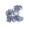
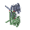
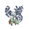
| Title | Structure of a bacterial ALDH16 active site mutant C295A complexed with p-nitrophenylacetate | ||||||
 Components Components | Aldehyde dehydrogenase | ||||||
 Keywords Keywords | OXIDOREDUCTASE / ALDEHYDE DEHYDROGENASE / ALDH16 / NAD / ROSSMANN FOLD | ||||||
| Function / homology | Aldehyde dehydrogenase / oxidoreductase activity, acting on the aldehyde or oxo group of donors, NAD or NADP as acceptor / Aldehyde dehydrogenase domain / Aldehyde dehydrogenase family / Aldehyde dehydrogenase, N-terminal / Aldehyde dehydrogenase, C-terminal / Aldehyde/histidinol dehydrogenase / 4-nitrophenyl acetate / Aldehyde dehydrogenase Function and homology information Function and homology information | ||||||
| Biological species |  Loktanella sp. 3ANDIMAR09 (bacteria) Loktanella sp. 3ANDIMAR09 (bacteria) | ||||||
| Method | X-RAY DIFFRACTION / SYNCHROTRON / MOLECULAR REPLACEMENT / Resolution: 1.488 Å | ||||||
 Authors Authors | Tanner, J.J. / Liu, L. | ||||||
| Funding support |  United States, 1items United States, 1items
| ||||||
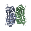
 Citation Citation | Journal: J Mol Biol / Year: 2019 Title: Crystal Structure of Aldehyde Dehydrogenase 16 Reveals Trans-Hierarchical Structural Similarity and a New Dimer. Authors: Li-Kai Liu / John J Tanner / Abstract: The aldehyde dehydrogenase (ALDH) superfamily is a vast group of enzymes that catalyze the NAD-dependent oxidation of aldehydes to carboxylic acids. ALDH16 is perhaps the most enigmatic member of the ...The aldehyde dehydrogenase (ALDH) superfamily is a vast group of enzymes that catalyze the NAD-dependent oxidation of aldehydes to carboxylic acids. ALDH16 is perhaps the most enigmatic member of the superfamily, owing to its extra C-terminal domain of unknown function and the absence of the essential catalytic cysteine residue in certain non-bacterial ALDH16 sequences. Herein we report the first production of recombinant ALDH16, the first biochemical characterization of ALDH16, and the first crystal structure of ALDH16. Recombinant expression systems were generated for the bacterial ALDH16 from Loktanella sp. and human ALDH16A1. Four high-resolution crystal structures of Loktanella ALDH16 were determined. Loktanella ALDH16 is found to be a bona fide enzyme, exhibiting NAD-binding, ALDH activity, and esterase activity. In contrast, human ALDH16A1 apparently lacks measurable aldehyde oxidation activity, suggesting that it is a pseudoenzyme, consistent with the absence of the catalytic Cys in its sequence. The fold of ALDH16 comprises three domains: NAD-binding, catalytic, and C-terminal. The latter is unique to ALDH16 and features a Rossmann fold connected to a protruding β-flap. The tertiary structural interactions of the C-terminal domain mimic the quaternary structural interactions of the classic ALDH superfamily dimer, a phenomenon we call "trans-hierarchical structural similarity." ALDH16 forms a unique dimer in solution, which mimics the classic ALDH superfamily dimer-of-dimer tetramer. Small-angle X-ray scattering shows that human ALDH16A1 has the same dimeric structure and fold as Loktanella ALDH16. We suggest that the Loktanella ALDH16 structure may be considered to be the archetype of the ALDH16 family. | ||||||
| History |
|
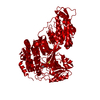
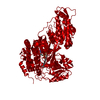
- Structure visualization
| Structure viewer | Molecule: MolmilJmol/JSmol |
|---|
- Downloads & links
-Download
| PDBx/mmCIF format | 6mvu.cif.gz | 571.1 KB | Display | PDBx/mmCIF format |
|---|---|---|---|---|
| PDB format | pdb6mvu.ent.gz | 465.3 KB | Display | PDB format |
| PDBx/mmJSON format | 6mvu.json.gz | Tree view | PDBx/mmJSON format | |
| Others |  Other downloads Other downloads |
-Validation report
| Arichive directory | https://data.pdbj.org/pub/pdb/validation_reports/mv/6mvuftp://data.pdbj.org/pub/pdb/validation_reports/mv/6mvu | HTTPS FTP |
|---|
-Related structure data
| Related structure data |  6mvrC  6mvsC  6mvtC 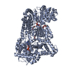 5kf6S S: Starting model for refinement C: citing same article ( |
|---|---|
| Similar structure data |


-Links
 PDBj
PDBj

- Assembly
| Deposited unit | 
| ||||||||
|---|---|---|---|---|---|---|---|---|---|
| 1 |
| ||||||||
| Unit cell |
|
-Components
| #1: Protein | Mass: 80842.977 Da / Num. of mol.: 2 / Mutation: C295A Source method: isolated from a genetically manipulated source Source: (gene. exp.) Loktanella sp. 3ANDIMAR09 (bacteria) / Gene: AN189_10795 / Production host: Escherichia coli (E. coli) / References: UniProt: A0A0Q3EUQ3#2: Chemical |   Mass: 181.145 Da / Num. of mol.: 2 / Source method: obtained synthetically / Formula: C8H7NO4 Mass: 181.145 Da / Num. of mol.: 2 / Source method: obtained synthetically / Formula: C8H7NO4#3: Chemical | Glycerol  Mass: 92.094 Da / Num. of mol.: 2 / Source method: obtained synthetically / Formula: C3H8O3 Mass: 92.094 Da / Num. of mol.: 2 / Source method: obtained synthetically / Formula: C3H8O3#4: Chemical | ChemComp-SO4 / Sulfate  Mass: 96.063 Da / Num. of mol.: 7 / Source method: obtained synthetically / Formula: SO4 Mass: 96.063 Da / Num. of mol.: 7 / Source method: obtained synthetically / Formula: SO4#5: Water | ChemComp-HOH / | Water Mass: 18.015 Da / Num. of mol.: 1224 / Source method: isolated from a natural source / Formula: H2O Mass: 18.015 Da / Num. of mol.: 1224 / Source method: isolated from a natural source / Formula: H2O |
|---|
-Experimental details
-Experiment
| Experiment | Method: X-RAY DIFFRACTION / Number of used crystals: 1 |
|---|
- Sample preparation
| Crystal | Density Matthews: 2.33 Å3/Da / Density % sol: 47.2 % |
|---|---|
| Crystal grow | Temperature: 293 K / Method: vapor diffusion, hanging drop / pH: 5.5 Details: Protein was concentrated to 6 mg/ml in a storage buffer consisting of 20 mM Tris-HCl at pH 8.0, 100 mM NaCl, 2.5% glycerol and 0.5 mM TCEP. The crystallization reservoir solution contained ...Details: Protein was concentrated to 6 mg/ml in a storage buffer consisting of 20 mM Tris-HCl at pH 8.0, 100 mM NaCl, 2.5% glycerol and 0.5 mM TCEP. The crystallization reservoir solution contained 20% (w/v) polyethylene glycol (PEG) 3350, 200 mM ammonium sulfate and 100 mM Bis-Tris at pH 5.5. |
-Data collection
| Diffraction | Mean temperature: 100 K / Serial crystal experiment: N |
|---|---|
| Diffraction source | Source: SYNCHROTRON / Site: APS / Beamline: 24-ID-C / Wavelength: 0.9791 Å |
| Detector | Type: DECTRIS PILATUS3 S 6M / Detector: PIXEL / Date: Nov 2, 2017 |
| Radiation | Protocol: SINGLE WAVELENGTH / Monochromatic (M) / Laue (L): M / Scattering type: x-ray |
| Radiation wavelength | Wavelength: 0.9791 Å / Relative weight: 1 |
| Reflection | Resolution: 1.488→119.62 Å / Num. obs: 470410 / % possible obs: 98.8 % / Redundancy: 6.7 % / Biso Wilson estimate: 17.19 Å2 / CC1/2: 0.998 / Rmerge(I) obs: 0.133 / Rpim(I) all: 0.055 / Rrim(I) all: 0.144 / Net I/σ(I): 8.8 |
| Reflection shell | Resolution: 1.488→1.51 Å / Redundancy: 6.5 % / Rmerge(I) obs: 1.799 / Num. unique obs: 11001 / CC1/2: 0.44 / Rpim(I) all: 0.748 / Rrim(I) all: 1.952 / % possible all: 90.8 |
- Processing
| Software |
| |||||||||||||||||||||||||||||||||||||||||||||||||||||||||||||||||||||||||||||||||||||||||||||||||||||||||||||||||||||||||||||||||||||||||||||||||||||||||||||||||||||||||||||||||||||||||||||||||||||||||||||||||||||||||
|---|---|---|---|---|---|---|---|---|---|---|---|---|---|---|---|---|---|---|---|---|---|---|---|---|---|---|---|---|---|---|---|---|---|---|---|---|---|---|---|---|---|---|---|---|---|---|---|---|---|---|---|---|---|---|---|---|---|---|---|---|---|---|---|---|---|---|---|---|---|---|---|---|---|---|---|---|---|---|---|---|---|---|---|---|---|---|---|---|---|---|---|---|---|---|---|---|---|---|---|---|---|---|---|---|---|---|---|---|---|---|---|---|---|---|---|---|---|---|---|---|---|---|---|---|---|---|---|---|---|---|---|---|---|---|---|---|---|---|---|---|---|---|---|---|---|---|---|---|---|---|---|---|---|---|---|---|---|---|---|---|---|---|---|---|---|---|---|---|---|---|---|---|---|---|---|---|---|---|---|---|---|---|---|---|---|---|---|---|---|---|---|---|---|---|---|---|---|---|---|---|---|---|---|---|---|---|---|---|---|---|---|---|---|---|---|---|---|---|
| Refinement | Method to determine structure: MOLECULAR REPLACEMENT Starting model: homology model built with Swiss-Model using 5kf6 as the template Resolution: 1.488→95.519 Å / SU ML: 0.21 / Cross valid method: THROUGHOUT / σ(F): 0.94 / Phase error: 23.68
| |||||||||||||||||||||||||||||||||||||||||||||||||||||||||||||||||||||||||||||||||||||||||||||||||||||||||||||||||||||||||||||||||||||||||||||||||||||||||||||||||||||||||||||||||||||||||||||||||||||||||||||||||||||||||
| Solvent computation | Shrinkage radii: 0.9 Å / VDW probe radii: 1.11 Å | |||||||||||||||||||||||||||||||||||||||||||||||||||||||||||||||||||||||||||||||||||||||||||||||||||||||||||||||||||||||||||||||||||||||||||||||||||||||||||||||||||||||||||||||||||||||||||||||||||||||||||||||||||||||||
| Displacement parameters | Biso max: 73.04 Å2 / Biso mean: 21.4238 Å2 / Biso min: 10.99 Å2 | |||||||||||||||||||||||||||||||||||||||||||||||||||||||||||||||||||||||||||||||||||||||||||||||||||||||||||||||||||||||||||||||||||||||||||||||||||||||||||||||||||||||||||||||||||||||||||||||||||||||||||||||||||||||||
| Refinement step | Cycle: final / Resolution: 1.488→95.519 Å
| |||||||||||||||||||||||||||||||||||||||||||||||||||||||||||||||||||||||||||||||||||||||||||||||||||||||||||||||||||||||||||||||||||||||||||||||||||||||||||||||||||||||||||||||||||||||||||||||||||||||||||||||||||||||||
| LS refinement shell | Refine-ID: X-RAY DIFFRACTION / Rfactor Rfree error: 0 / Total num. of bins used: 30
| |||||||||||||||||||||||||||||||||||||||||||||||||||||||||||||||||||||||||||||||||||||||||||||||||||||||||||||||||||||||||||||||||||||||||||||||||||||||||||||||||||||||||||||||||||||||||||||||||||||||||||||||||||||||||
| Refinement TLS params. | Method: refined / Refine-ID: X-RAY DIFFRACTION
| |||||||||||||||||||||||||||||||||||||||||||||||||||||||||||||||||||||||||||||||||||||||||||||||||||||||||||||||||||||||||||||||||||||||||||||||||||||||||||||||||||||||||||||||||||||||||||||||||||||||||||||||||||||||||
| Refinement TLS group |
|
