 Movie
Movie Controller

[English] 日本語
 Yorodumi
- PDB-6mtk: Crystal structure of Tryptophanyl-tRNA synthetase from Elizabethk... -
+ Open data

- Basic information
| Entry | Database: PDB / ID: 6mtk | ||||||
|---|---|---|---|---|---|---|---|
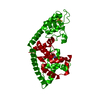
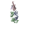
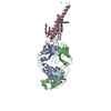
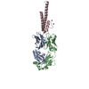
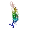
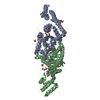
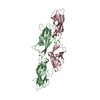
| Title | Crystal structure of Tryptophanyl-tRNA synthetase from Elizabethkingia anophelis NUHP1 | ||||||
 Components Components | Tryptophanyl-tRNA synthetase | ||||||
 Keywords Keywords |  LIGASE / SSGCID / Structural Genomics / Elizabethkingia anophelis / BD94_0195 / Tryptophanyl-tRNA synthetase / Seattle Structural Genomics Center for Infectious Disease LIGASE / SSGCID / Structural Genomics / Elizabethkingia anophelis / BD94_0195 / Tryptophanyl-tRNA synthetase / Seattle Structural Genomics Center for Infectious Disease | ||||||
| Function / homology |  Function and homology informationtryptophan-tRNA ligase / tryptophan-tRNA ligase activity / tryptophanyl-tRNA aminoacylation / ATP binding Function and homology informationtryptophan-tRNA ligase / tryptophan-tRNA ligase activity / tryptophanyl-tRNA aminoacylation / ATP bindingSimilarity search - Function | ||||||
| Biological species |  Elizabethkingia anophelis NUHP1 (bacteria) Elizabethkingia anophelis NUHP1 (bacteria) | ||||||
| Method | X-RAY DIFFRACTION / MOLECULAR REPLACEMENT / Resolution: 2 Å | ||||||
 Authors Authors | Seattle Structural Genomics Center for Infectious Disease (SSGCID) | ||||||
 Citation Citation | Journal: To be Published Title: Crystal structure of Tryptophanyl-tRNA synthetase from Elizabethkingia anophelis NUHP1 Authors: Abendroth, J. / Dranow, D.M. / Lorimer, D.D. / Horanyi, P.S. / Edwards, T.E. | ||||||
| History |
|
- Structure visualization
| Structure viewer | Molecule: MolmilJmol/JSmol |
|---|
- Downloads & links
-Download
| PDBx/mmCIF format | 6mtk.cif.gz | 141.1 KB | Display | PDBx/mmCIF format |
|---|---|---|---|---|
| PDB format | pdb6mtk.ent.gz | 108.2 KB | Display | PDB format |
| PDBx/mmJSON format | 6mtk.json.gz | Tree view | PDBx/mmJSON format | |
| Others |  Other downloads Other downloads |
-Validation report
| Arichive directory | https://data.pdbj.org/pub/pdb/validation_reports/mt/6mtkftp://data.pdbj.org/pub/pdb/validation_reports/mt/6mtk | HTTPS FTP |
|---|
-Related structure data
| Related structure data |  3tzlS S: Starting model for refinement |
|---|---|
| Similar structure data | |
| Other databases |


-Links
 PDBj
PDBj- Assembly
| Deposited unit | 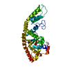
| |||||||||
|---|---|---|---|---|---|---|---|---|---|---|
| 1 | 
| |||||||||
| Unit cell |
| |||||||||
| Components on special symmetry positions |
|
-Components
| #1: Protein | Mass: 37500.879 Da / Num. of mol.: 1 Source method: isolated from a genetically manipulated source Source: (gene. exp.) Elizabethkingia anophelis NUHP1 (bacteria)Gene: BD94_0195 / Plasmid: ElanA.00743.a.B1 Production host: Escherichia coli 'BL21-Gold(DE3)pLysS AG' (bacteria)Strain (production host): BL21(DE3) / References: UniProt: A0A077EER2, tryptophan-tRNA ligase |
|---|---|
| #2: Water | ChemComp-HOH / Water Mass: 18.015 Da / Num. of mol.: 123 / Source method: isolated from a natural source / Formula: H2O Mass: 18.015 Da / Num. of mol.: 123 / Source method: isolated from a natural source / Formula: H2O |
-Experimental details
-Experiment
| Experiment | Method: X-RAY DIFFRACTION / Number of used crystals: 1 |
|---|
- Sample preparation
| Crystal | Density Matthews: 2.17 Å3/Da / Density % sol: 43.3 % |
|---|---|
| Crystal grow | Temperature: 290 K / Method: vapor diffusion, sitting drop / pH: 8.5 Details: 22.6 mg/mL ElanA.00743.a.B1.PS38400 + Molecular Dimensions Morpheus screen, condition C10 (10% w/v PEG8000, 20% v/v ethylene glycol, 30 mM each NPS, 100 mM bicine/Trizma base, pH 8.5, sodium ...Details: 22.6 mg/mL ElanA.00743.a.B1.PS38400 + Molecular Dimensions Morpheus screen, condition C10 (10% w/v PEG8000, 20% v/v ethylene glycol, 30 mM each NPS, 100 mM bicine/Trizma base, pH 8.5, sodium nitrate, 0.3 M disodium hydrogen phosphate, 0.3 M ammonium sulfate), cryoprotectant: direct, Tray 299155c10, puck OVZ1-7 |
-Data collection
| Diffraction | Mean temperature: 100 K / Serial crystal experiment: N | ||||||||||||||||||||||||||||||||||||||||||||||||||||||||||||||||||||||||||||||||||||||||||||||||||||||||||||||||||||||||||||||||||||||||||||||||||||||||||||||||||||||||
|---|---|---|---|---|---|---|---|---|---|---|---|---|---|---|---|---|---|---|---|---|---|---|---|---|---|---|---|---|---|---|---|---|---|---|---|---|---|---|---|---|---|---|---|---|---|---|---|---|---|---|---|---|---|---|---|---|---|---|---|---|---|---|---|---|---|---|---|---|---|---|---|---|---|---|---|---|---|---|---|---|---|---|---|---|---|---|---|---|---|---|---|---|---|---|---|---|---|---|---|---|---|---|---|---|---|---|---|---|---|---|---|---|---|---|---|---|---|---|---|---|---|---|---|---|---|---|---|---|---|---|---|---|---|---|---|---|---|---|---|---|---|---|---|---|---|---|---|---|---|---|---|---|---|---|---|---|---|---|---|---|---|---|---|---|---|---|---|---|---|
| Diffraction source | Source: ROTATING ANODE / Type: RIGAKU FR-E+ SUPERBRIGHT / Wavelength: 1.5418 Å | ||||||||||||||||||||||||||||||||||||||||||||||||||||||||||||||||||||||||||||||||||||||||||||||||||||||||||||||||||||||||||||||||||||||||||||||||||||||||||||||||||||||||
| Detector | Type: RIGAKU SATURN 944+ / Detector: CCD / Date: Sep 14, 2018 | ||||||||||||||||||||||||||||||||||||||||||||||||||||||||||||||||||||||||||||||||||||||||||||||||||||||||||||||||||||||||||||||||||||||||||||||||||||||||||||||||||||||||
| Radiation | Monochromator: RIGAKU VARIMAX / Protocol: SINGLE WAVELENGTH / Monochromatic (M) / Laue (L): M / Scattering type: x-ray | ||||||||||||||||||||||||||||||||||||||||||||||||||||||||||||||||||||||||||||||||||||||||||||||||||||||||||||||||||||||||||||||||||||||||||||||||||||||||||||||||||||||||
| Radiation wavelength | Wavelength: 1.5418 Å / Relative weight: 1 | ||||||||||||||||||||||||||||||||||||||||||||||||||||||||||||||||||||||||||||||||||||||||||||||||||||||||||||||||||||||||||||||||||||||||||||||||||||||||||||||||||||||||
| Reflection | Resolution: 2→47.095 Å / Num. obs: 22221 / % possible obs: 99.3 % / Redundancy: 12.517 % / Biso Wilson estimate: 45.669 Å2 / CC1/2: 0.999 / Rmerge(I) obs: 0.041 / Rrim(I) all: 0.043 / Χ2: 1.031 / Net I/σ(I): 33.22 | ||||||||||||||||||||||||||||||||||||||||||||||||||||||||||||||||||||||||||||||||||||||||||||||||||||||||||||||||||||||||||||||||||||||||||||||||||||||||||||||||||||||||
| Reflection shell | Diffraction-ID: 1
|
- Processing
| Software |
| |||||||||||||||||||||||||||||||||||||||||||||||||||||||||||||||||||||||||||||||||||||||||||||||||||||||||
|---|---|---|---|---|---|---|---|---|---|---|---|---|---|---|---|---|---|---|---|---|---|---|---|---|---|---|---|---|---|---|---|---|---|---|---|---|---|---|---|---|---|---|---|---|---|---|---|---|---|---|---|---|---|---|---|---|---|---|---|---|---|---|---|---|---|---|---|---|---|---|---|---|---|---|---|---|---|---|---|---|---|---|---|---|---|---|---|---|---|---|---|---|---|---|---|---|---|---|---|---|---|---|---|---|---|---|
| Refinement | Method to determine structure: MOLECULAR REPLACEMENT Starting model: PDB entry 3TZL as per MoRDa Resolution: 2→47.095 Å / SU ML: 0.27 / Cross valid method: THROUGHOUT / σ(F): 1.34 / Phase error: 31.03
| |||||||||||||||||||||||||||||||||||||||||||||||||||||||||||||||||||||||||||||||||||||||||||||||||||||||||
| Solvent computation | Shrinkage radii: 0.9 Å / VDW probe radii: 1.11 Å | |||||||||||||||||||||||||||||||||||||||||||||||||||||||||||||||||||||||||||||||||||||||||||||||||||||||||
| Displacement parameters | Biso max: 115.07 Å2 / Biso mean: 50.5227 Å2 / Biso min: 23.75 Å2 | |||||||||||||||||||||||||||||||||||||||||||||||||||||||||||||||||||||||||||||||||||||||||||||||||||||||||
| Refinement step | Cycle: final / Resolution: 2→47.095 Å
| |||||||||||||||||||||||||||||||||||||||||||||||||||||||||||||||||||||||||||||||||||||||||||||||||||||||||
| Refine LS restraints |
| |||||||||||||||||||||||||||||||||||||||||||||||||||||||||||||||||||||||||||||||||||||||||||||||||||||||||
| LS refinement shell | Refine-ID: X-RAY DIFFRACTION / Rfactor Rfree error: 0 / Total num. of bins used: 14
| |||||||||||||||||||||||||||||||||||||||||||||||||||||||||||||||||||||||||||||||||||||||||||||||||||||||||
| Refinement TLS params. | Method: refined / Refine-ID: X-RAY DIFFRACTION
| |||||||||||||||||||||||||||||||||||||||||||||||||||||||||||||||||||||||||||||||||||||||||||||||||||||||||
| Refinement TLS group |
|
