 Movie
Movie Controller


+ Open data

- Basic information
| Entry | Database: PDB / ID: 4xkv | |||||||||
|---|---|---|---|---|---|---|---|---|---|---|
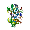
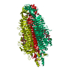
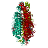
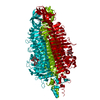
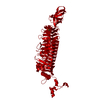
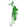
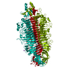
| Title | Tailspike protein mutant D339N of E. coli bacteriophage HK620 | |||||||||
 Components Components | Tail spike protein | |||||||||
 Keywords Keywords |  VIRAL PROTEIN / beta helix / protein-carbohydrate complex / pectin lyase fold / metal binding protein VIRAL PROTEIN / beta helix / protein-carbohydrate complex / pectin lyase fold / metal binding protein | |||||||||
| Function / homology |  Function and homology information Function and homology informationbiological process involved in interaction with host / viral life cycle / virion componentSimilarity search - Function | |||||||||
| Biological species |  Salmonella phage HK620 (virus) Salmonella phage HK620 (virus) | |||||||||
| Method | X-RAY DIFFRACTION / SYNCHROTRON / MOLECULAR REPLACEMENT / Resolution: 2.1 Å | |||||||||
 Authors Authors | Gohlke, U. / Broeker, N.K. / Heinemann, U. / Seckler, R. / Barbirz, S. | |||||||||
| Funding support |  Germany, 1items Germany, 1items
| |||||||||
 Citation Citation | Journal: to be published Title: Enthalpic cost of water removal from a hydrophobic glucose binding cavity on HK620 tailspike protein. Authors: Gohlke, U. / Broeker, N.K. / Kunstmann, S. / Santer, M. / Heinemann, U. / Lipowski, R. / Seckler, R. / Barbirz, S. #1: Journal: Glycobiology / Year: 2013Title: Single amino acid exchange in bacteriophage HK620 tailspike protein results in thousand-fold increase of its oligosaccharide affinity. Authors: Broeker, N.K. / Gohlke, U. / Mueller, J.J. / Uetrecht, C. / Heinemann, U. / Seckler, R. / Barbirz, S. | |||||||||
| History |
|
- Structure visualization
| Structure viewer | Molecule: MolmilJmol/JSmol |
|---|
- Downloads & links
-Download
| PDBx/mmCIF format | 4xkv.cif.gz | 237.2 KB | Display | PDBx/mmCIF format |
|---|---|---|---|---|
| PDB format | pdb4xkv.ent.gz | 189.8 KB | Display | PDB format |
| PDBx/mmJSON format | 4xkv.json.gz | Tree view | PDBx/mmJSON format | |
| Others |  Other downloads Other downloads |
-Validation report
| Arichive directory | https://data.pdbj.org/pub/pdb/validation_reports/xk/4xkvftp://data.pdbj.org/pub/pdb/validation_reports/xk/4xkv | HTTPS FTP |
|---|
-Related structure data
| Related structure data |  4xkwC 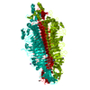 4xl9C  4xlaC  4xlcC  4xleC  4xlfC 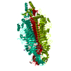 4xlhC 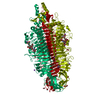 4xm3SC  4xmyC 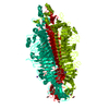 4xn0C 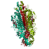 4xn3C 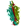 4xnfC 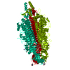 4xonC  4xopC 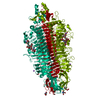 4xorC 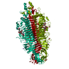 4xotC  4xqfC  4xr6C  4yejC  4yelC  4pat S: Starting model for refinement C: citing same article ( |
|---|---|
| Similar structure data |


-Links
 PDBj
PDBj- Assembly
| Deposited unit | 
| |||||||||
|---|---|---|---|---|---|---|---|---|---|---|
| 1 | 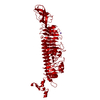
| |||||||||
| Unit cell |
| |||||||||
| Components on special symmetry positions |
| |||||||||
| Details | The biological assembly is a trimer generated from the monomer in the asymmetric unit by the operations: -y+1,x-y,z and -x+y+1,-x+1,z. |
-Components
| #1: Protein | Mass: 64248.957 Da / Num. of mol.: 1 / Fragment: head binding, UNP residues 1-596 / Mutation: D339N Source method: isolated from a genetically manipulated source Source: (gene. exp.) Salmonella phage HK620 (virus) / Plasmid: pET11d / Production host:  Escherichia coli (E. coli) / Strain (production host): BL21 / References: UniProt: Q9AYY6 Escherichia coli (E. coli) / Strain (production host): BL21 / References: UniProt: Q9AYY6 | ||||
|---|---|---|---|---|---|
| #2: Chemical | ChemComp-TRS / Tris  Mass: 122.143 Da / Num. of mol.: 1 / Source method: obtained synthetically / Formula: C4H12NO3 / Comment: pH buffer*YM Mass: 122.143 Da / Num. of mol.: 1 / Source method: obtained synthetically / Formula: C4H12NO3 / Comment: pH buffer*YM | ||||
| #3: Chemical | Formic acid  Mass: 46.025 Da / Num. of mol.: 3 / Source method: obtained synthetically / Formula: CH2O2 Mass: 46.025 Da / Num. of mol.: 3 / Source method: obtained synthetically / Formula: CH2O2#4: Chemical | ChemComp-NA / |   Mass: 22.990 Da / Num. of mol.: 1 / Source method: obtained synthetically / Formula: Na Mass: 22.990 Da / Num. of mol.: 1 / Source method: obtained synthetically / Formula: Na#5: Water | ChemComp-HOH / | Water Mass: 18.015 Da / Num. of mol.: 262 / Source method: isolated from a natural source / Formula: H2O Mass: 18.015 Da / Num. of mol.: 262 / Source method: isolated from a natural source / Formula: H2O |
-Experimental details
-Experiment
| Experiment | Method: X-RAY DIFFRACTION / Number of used crystals: 1 |
|---|
- Sample preparation
| Crystal | Density Matthews: 2.14 Å3/Da / Density % sol: 42.57 % |
|---|---|
| Crystal grow | Temperature: 298 K / Method: vapor diffusion, hanging drop / pH: 8.5 / Details: 0.1 M Tris-HCl, 3.5 M Sodium formate |
-Data collection
| Diffraction | Mean temperature: 100 K | |||||||||||||||||||||||||||
|---|---|---|---|---|---|---|---|---|---|---|---|---|---|---|---|---|---|---|---|---|---|---|---|---|---|---|---|---|
| Diffraction source | Source: SYNCHROTRON / Site: BESSY / Beamline: 14.1 / Wavelength: 0.91841 Å | |||||||||||||||||||||||||||
| Detector | Type: DECTRIS PILATUS 6M / Detector: PIXEL / Date: Jan 28, 2014 / Details: mirrors | |||||||||||||||||||||||||||
| Radiation | Monochromator: SI-111 CRYSTAL / Protocol: SINGLE WAVELENGTH / Monochromatic (M) / Laue (L): M / Scattering type: x-ray | |||||||||||||||||||||||||||
| Radiation wavelength | Wavelength: 0.91841 Å / Relative weight: 1 | |||||||||||||||||||||||||||
| Reflection | Resolution: 2.1→43.05 Å / Num. obs: 26540 / % possible obs: 80.1 % / Redundancy: 3.5 % / CC1/2: 0.995 / Rmerge(I) obs: 0.08 / Rpim(I) all: 0.043 / Net I/σ(I): 10.1 / Num. measured all: 92726 | |||||||||||||||||||||||||||
| Reflection shell | Diffraction-ID: 1 / Rejects: 0
|
- Processing
| Software |
| |||||||||||||||||||||||||||||||||||||||||||||||||||||||||||||||||||||||||||||||||||||||||||||||||||||||||||||||||||||||||||||||||||||||||||||||||||||||||||||||||||||||||||||||||||||||||||||||||||||||||||||||||||||||||||||||||
|---|---|---|---|---|---|---|---|---|---|---|---|---|---|---|---|---|---|---|---|---|---|---|---|---|---|---|---|---|---|---|---|---|---|---|---|---|---|---|---|---|---|---|---|---|---|---|---|---|---|---|---|---|---|---|---|---|---|---|---|---|---|---|---|---|---|---|---|---|---|---|---|---|---|---|---|---|---|---|---|---|---|---|---|---|---|---|---|---|---|---|---|---|---|---|---|---|---|---|---|---|---|---|---|---|---|---|---|---|---|---|---|---|---|---|---|---|---|---|---|---|---|---|---|---|---|---|---|---|---|---|---|---|---|---|---|---|---|---|---|---|---|---|---|---|---|---|---|---|---|---|---|---|---|---|---|---|---|---|---|---|---|---|---|---|---|---|---|---|---|---|---|---|---|---|---|---|---|---|---|---|---|---|---|---|---|---|---|---|---|---|---|---|---|---|---|---|---|---|---|---|---|---|---|---|---|---|---|---|---|---|---|---|---|---|---|---|---|---|---|---|---|---|---|---|---|---|
| Refinement | Method to determine structure: MOLECULAR REPLACEMENT Starting model: 4xm3 Resolution: 2.1→43.05 Å / Cor.coef. Fo:Fc: 0.92 / Cor.coef. Fo:Fc free: 0.871 / WRfactor Rfree: 0.2727 / WRfactor Rwork: 0.2182 / FOM work R set: 0.7506 / SU B: 15.096 / SU ML: 0.209 / SU R Cruickshank DPI: 0.4765 / SU Rfree: 0.2845 / Cross valid method: THROUGHOUT / σ(F): 0 / ESU R: 0.477 / ESU R Free: 0.284 / Stereochemistry target values: MAXIMUM LIKELIHOOD Details: HYDROGENS HAVE BEEN ADDED IN THE RIDING POSITIONS U VALUES : WITH TLS ADDED
| |||||||||||||||||||||||||||||||||||||||||||||||||||||||||||||||||||||||||||||||||||||||||||||||||||||||||||||||||||||||||||||||||||||||||||||||||||||||||||||||||||||||||||||||||||||||||||||||||||||||||||||||||||||||||||||||||
| Solvent computation | Ion probe radii: 0.8 Å / Shrinkage radii: 0.8 Å / VDW probe radii: 1.2 Å / Solvent model: MASK | |||||||||||||||||||||||||||||||||||||||||||||||||||||||||||||||||||||||||||||||||||||||||||||||||||||||||||||||||||||||||||||||||||||||||||||||||||||||||||||||||||||||||||||||||||||||||||||||||||||||||||||||||||||||||||||||||
| Displacement parameters | Biso max: 56.45 Å2 / Biso mean: 22.591 Å2 / Biso min: 7.48 Å2
| |||||||||||||||||||||||||||||||||||||||||||||||||||||||||||||||||||||||||||||||||||||||||||||||||||||||||||||||||||||||||||||||||||||||||||||||||||||||||||||||||||||||||||||||||||||||||||||||||||||||||||||||||||||||||||||||||
| Refinement step | Cycle: final / Resolution: 2.1→43.05 Å
| |||||||||||||||||||||||||||||||||||||||||||||||||||||||||||||||||||||||||||||||||||||||||||||||||||||||||||||||||||||||||||||||||||||||||||||||||||||||||||||||||||||||||||||||||||||||||||||||||||||||||||||||||||||||||||||||||
| Refine LS restraints |
| |||||||||||||||||||||||||||||||||||||||||||||||||||||||||||||||||||||||||||||||||||||||||||||||||||||||||||||||||||||||||||||||||||||||||||||||||||||||||||||||||||||||||||||||||||||||||||||||||||||||||||||||||||||||||||||||||
| LS refinement shell | Resolution: 2.1→2.155 Å / Total num. of bins used: 20
| |||||||||||||||||||||||||||||||||||||||||||||||||||||||||||||||||||||||||||||||||||||||||||||||||||||||||||||||||||||||||||||||||||||||||||||||||||||||||||||||||||||||||||||||||||||||||||||||||||||||||||||||||||||||||||||||||
| Refinement TLS params. | Method: refined / Refine-ID: X-RAY DIFFRACTION
| |||||||||||||||||||||||||||||||||||||||||||||||||||||||||||||||||||||||||||||||||||||||||||||||||||||||||||||||||||||||||||||||||||||||||||||||||||||||||||||||||||||||||||||||||||||||||||||||||||||||||||||||||||||||||||||||||
| Refinement TLS group |
|
