 Movie
Movie Controller

[English] 日本語
 Yorodumi
- PDB-6mny: Crystal structure of mouse BTK kinase domain in complex with comp... -
+ Open data

- Basic information
| Entry | Database: PDB / ID: 6mny | ||||||
|---|---|---|---|---|---|---|---|
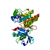
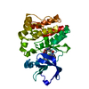
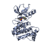
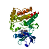
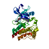
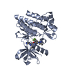
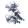
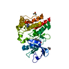
| Title | Crystal structure of mouse BTK kinase domain in complex with compound 9a | ||||||
 Components Components | Tyrosine-protein kinase | ||||||
 Keywords Keywords |  TRANSFERASE / kinase / drug design TRANSFERASE / kinase / drug design | ||||||
| Function / homology |  Function and homology informationtransmembrane receptor protein tyrosine kinase activity / non-specific protein-tyrosine kinase / non-membrane spanning protein tyrosine kinase activity / adaptive immune response / intracellular signal transduction / ATP binding / metal ion binding Function and homology informationtransmembrane receptor protein tyrosine kinase activity / non-specific protein-tyrosine kinase / non-membrane spanning protein tyrosine kinase activity / adaptive immune response / intracellular signal transduction / ATP binding / metal ion bindingSimilarity search - Function | ||||||
| Biological species |  Mus musculus (house mouse) Mus musculus (house mouse) | ||||||
| Method | X-RAY DIFFRACTION / SYNCHROTRON / Resolution: 2.8 Å | ||||||
 Authors Authors | Han, S. / Caspers, N. / Ohren, J.O. | ||||||
 Citation Citation | Journal: ACS Med Chem Lett / Year: 2019 Title: Aminopyrazole Carboxamide Bruton's Tyrosine Kinase Inhibitors. Irreversible to Reversible Covalent Reactive Group Tuning. Authors: Schnute, M.E. / Benoit, S.E. / Buchler, I.P. / Caspers, N. / Grapperhaus, M.L. / Han, S. / Hotchandani, R. / Huang, N. / Hughes, R.O. / Juba, B.M. / Kim, K.H. / Liu, E. / McCarthy, E. / ...Authors: Schnute, M.E. / Benoit, S.E. / Buchler, I.P. / Caspers, N. / Grapperhaus, M.L. / Han, S. / Hotchandani, R. / Huang, N. / Hughes, R.O. / Juba, B.M. / Kim, K.H. / Liu, E. / McCarthy, E. / Messing, D. / Miyashiro, J.S. / Mohan, S. / O'Connell, T.N. / Ohren, J.F. / Parikh, M.D. / Schmidt, M. / Selness, S.R. / Springer, J.R. / Thanabal, V. / Trujillo, J.I. / Walker, D.P. / Wan, Z.K. / Withka, J.M. / Wittwer, A.J. / Wood, N.L. / Xing, L. / Zapf, C.W. / Douhan III, J. | ||||||
| History |
|
- Structure visualization
| Structure viewer | Molecule: MolmilJmol/JSmol |
|---|
- Downloads & links
-Download
| PDBx/mmCIF format | 6mny.cif.gz | 218.8 KB | Display | PDBx/mmCIF format |
|---|---|---|---|---|
| PDB format | pdb6mny.ent.gz | 181.9 KB | Display | PDB format |
| PDBx/mmJSON format | 6mny.json.gz | Tree view | PDBx/mmJSON format | |
| Others |  Other downloads Other downloads |
-Validation report
| Arichive directory | https://data.pdbj.org/pub/pdb/validation_reports/mn/6mnyftp://data.pdbj.org/pub/pdb/validation_reports/mn/6mny | HTTPS FTP |
|---|
-Related structure data
| Similar structure data |
|---|


-Links
 PDBj
PDBj
- Assembly
| Deposited unit | 
| ||||||||
|---|---|---|---|---|---|---|---|---|---|
| 1 | 
| ||||||||
| 2 | 
| ||||||||
| Unit cell |
|
-Components
| #1: Protein | Mass: 32037.719 Da / Num. of mol.: 2 / Fragment: Protein kinase domain, residues 384-659 Source method: isolated from a genetically manipulated source Source: (gene. exp.) Mus musculus (house mouse) / Gene: Btk / Production host:   Spodoptera frugiperda (fall armyworm) Spodoptera frugiperda (fall armyworm)References: UniProt: Q7TMU1, non-specific protein-tyrosine kinase#2: Chemical | ChemComp-JVP / |   Mass: 438.430 Da / Num. of mol.: 1 / Source method: obtained synthetically / Formula: C22H20F2N6O2 Mass: 438.430 Da / Num. of mol.: 1 / Source method: obtained synthetically / Formula: C22H20F2N6O2#3: Water | ChemComp-HOH / | Water Mass: 18.015 Da / Num. of mol.: 148 / Source method: isolated from a natural source / Formula: H2O Mass: 18.015 Da / Num. of mol.: 148 / Source method: isolated from a natural source / Formula: H2O |
|---|
-Experimental details
-Experiment
| Experiment | Method: X-RAY DIFFRACTION / Number of used crystals: 1 |
|---|
- Sample preparation
| Crystal | Density Matthews: 2.51 Å3/Da / Density % sol: 50.99 % |
|---|---|
| Crystal grow | Temperature: 277 K / Method: vapor diffusion, hanging drop / pH: 6 Details: 12% PEG 3350, 0.1 M Bis-Tris pH 6.0, 0.1 M sodium malonate |
-Data collection
| Diffraction | Mean temperature: 173 K / Serial crystal experiment: N |
|---|---|
| Diffraction source | Source: SYNCHROTRON / Site: APS  / Beamline: 17-ID / Wavelength: 1 Å / Beamline: 17-ID / Wavelength: 1 Å |
| Detector | Type: DECTRIS PILATUS3 6M / Detector: PIXEL / Date: Dec 1, 2017 |
| Radiation | Protocol: SINGLE WAVELENGTH / Monochromatic (M) / Laue (L): M / Scattering type: x-ray |
| Radiation wavelength | Wavelength: 1 Å / Relative weight: 1 |
| Reflection | Resolution: 2.8→68.1 Å / Num. obs: 16257 / % possible obs: 96.8 % / Redundancy: 1.8 % / Biso Wilson estimate: 52.55 Å2 / Rmerge(I) obs: 0.147 / Net I/σ(I): 6.3 |
| Reflection shell | Resolution: 2.8→2.89 Å / Rmerge(I) obs: 0.467 / Num. unique obs: 2364 / % possible all: 96.1 |
- Processing
| Software |
| ||||||||||||||||||||||||||||||||||||||||||||||||||||||||||||||||||||||||||||||||||||||||||||||||||||||||||||||||||
|---|---|---|---|---|---|---|---|---|---|---|---|---|---|---|---|---|---|---|---|---|---|---|---|---|---|---|---|---|---|---|---|---|---|---|---|---|---|---|---|---|---|---|---|---|---|---|---|---|---|---|---|---|---|---|---|---|---|---|---|---|---|---|---|---|---|---|---|---|---|---|---|---|---|---|---|---|---|---|---|---|---|---|---|---|---|---|---|---|---|---|---|---|---|---|---|---|---|---|---|---|---|---|---|---|---|---|---|---|---|---|---|---|---|---|---|
| Refinement | Resolution: 2.8→68.1 Å / Cor.coef. Fo:Fc: 0.886 / Cor.coef. Fo:Fc free: 0.807 / Cross valid method: THROUGHOUT / σ(F): 0 / SU Rfree Blow DPI: 0.385
| ||||||||||||||||||||||||||||||||||||||||||||||||||||||||||||||||||||||||||||||||||||||||||||||||||||||||||||||||||
| Displacement parameters | Biso mean: 30.97 Å2
| ||||||||||||||||||||||||||||||||||||||||||||||||||||||||||||||||||||||||||||||||||||||||||||||||||||||||||||||||||
| Refine analyze | Luzzati coordinate error obs: 0.35 Å | ||||||||||||||||||||||||||||||||||||||||||||||||||||||||||||||||||||||||||||||||||||||||||||||||||||||||||||||||||
| Refinement step | Cycle: 1 / Resolution: 2.8→68.1 Å
| ||||||||||||||||||||||||||||||||||||||||||||||||||||||||||||||||||||||||||||||||||||||||||||||||||||||||||||||||||
| Refine LS restraints |
| ||||||||||||||||||||||||||||||||||||||||||||||||||||||||||||||||||||||||||||||||||||||||||||||||||||||||||||||||||
| LS refinement shell | Resolution: 2.8→3.02 Å / Total num. of bins used: 7
| ||||||||||||||||||||||||||||||||||||||||||||||||||||||||||||||||||||||||||||||||||||||||||||||||||||||||||||||||||
| Refinement TLS params. | Method: refined / Refine-ID: X-RAY DIFFRACTION
| ||||||||||||||||||||||||||||||||||||||||||||||||||||||||||||||||||||||||||||||||||||||||||||||||||||||||||||||||||
| Refinement TLS group |
|
