 Movie
Movie Controller

[English] 日本語
 Yorodumi
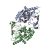
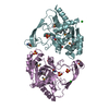
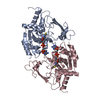
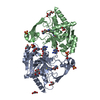
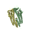
- PDB-6mn2: Crystal structure of meta-AAC0038, an environmental aminoglycosid... -
+ Open data

- Basic information
| Entry | Database: PDB / ID: 6mn2 | |||||||||
|---|---|---|---|---|---|---|---|---|---|---|
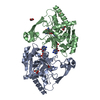
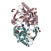
| Title | Crystal structure of meta-AAC0038, an environmental aminoglycoside resistance enzyme, mutant H168A in abortive complex with sisomicin-CoA | |||||||||
 Components Components | Aminoglycoside N(3)-acetyltransferase | |||||||||
 Keywords Keywords | TRANSFERASE/ANTIBIOTIC / ANTIBIOTIC_NAT FAMILY /  ACETYLTRANSFERASE / AMINOGLYCOSIDE / ANTIBIOTIC RESISTANCE / METAGENOME / SOIL / COENZYME A / sisomicin / CSGID / TRANSFERASE / CENTER FOR STRUCTURAL GENOMICS OF INFECTIOUS DISEASES / National Institute of Allergy and Infectious Diseases / NIAID / TRANSFERASE-ANTIBIOTIC complex ACETYLTRANSFERASE / AMINOGLYCOSIDE / ANTIBIOTIC RESISTANCE / METAGENOME / SOIL / COENZYME A / sisomicin / CSGID / TRANSFERASE / CENTER FOR STRUCTURAL GENOMICS OF INFECTIOUS DISEASES / National Institute of Allergy and Infectious Diseases / NIAID / TRANSFERASE-ANTIBIOTIC complex | |||||||||
| Function / homology |  Function and homology information Function and homology informationaminoglycoside 3-N-acetyltransferase / aminoglycoside 3-N-acetyltransferase activity / response to antibiotic Similarity search - Function | |||||||||
| Biological species |  uncultured bacterium (environmental samples) uncultured bacterium (environmental samples) | |||||||||
| Method | X-RAY DIFFRACTION / SYNCHROTRON / MOLECULAR REPLACEMENT / Resolution: 2.744 Å | |||||||||
 Authors Authors | Stogios, P.J. / Skarina, T. / Michalska, K. / Xu, Z. / Yim, V. / Savchenko, A. / Joachimiak, A. / Satchell, K.J. / Center for Structural Genomics of Infectious Diseases (CSGID) | |||||||||
| Funding support |  United States, 2items United States, 2items
| |||||||||
 Citation Citation | Journal: To Be Published Title: Crystal structure of meta-AAC0038, an environmental aminoglycoside resistance enzyme, mutant H168A in abortive complex with sisomicin-CoA Authors: Stogios, P.J. | |||||||||
| History |
|
- Structure visualization
| Structure viewer | Molecule: MolmilJmol/JSmol |
|---|
- Downloads & links
-Download
| PDBx/mmCIF format | 6mn2.cif.gz | 221.6 KB | Display | PDBx/mmCIF format |
|---|---|---|---|---|
| PDB format | pdb6mn2.ent.gz | 178.9 KB | Display | PDB format |
| PDBx/mmJSON format | 6mn2.json.gz | Tree view | PDBx/mmJSON format | |
| Others |  Other downloads Other downloads |
-Validation report
| Arichive directory | https://data.pdbj.org/pub/pdb/validation_reports/mn/6mn2ftp://data.pdbj.org/pub/pdb/validation_reports/mn/6mn2 | HTTPS FTP |
|---|
-Related structure data
| Related structure data | 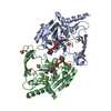 5ht0S S: Starting model for refinement |
|---|---|
| Similar structure data | |
| Other databases |


-Links
 PDBj
PDBj


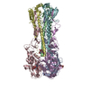
- Assembly
| Deposited unit | 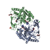
| ||||||||
|---|---|---|---|---|---|---|---|---|---|
| 1 |
| ||||||||
| Unit cell |
|
-Components
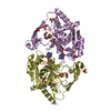
-Protein , 1 types, 2 molecules AB
| #1: Protein | Mass: 28654.350 Da / Num. of mol.: 2 / Mutation: H168A Source method: isolated from a genetically manipulated source Source: (gene. exp.) uncultured bacterium (environmental samples)Plasmid: pMCSG53 / Production host: Escherichia coli (E. coli) / Strain (production host): BL21(DE3)-MagicReferences: UniProt: A0A059X981, aminoglycoside 3-N-acetyltransferase |
|---|
-Non-polymers , 6 types, 130 molecules 
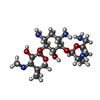


| #2: Chemical | Coenzyme A Mass: 767.534 Da / Num. of mol.: 2 / Source method: obtained synthetically / Formula: C21H36N7O16P3S Mass: 767.534 Da / Num. of mol.: 2 / Source method: obtained synthetically / Formula: C21H36N7O16P3S#3: Chemical | Sisomicin Mass: 447.526 Da / Num. of mol.: 2 / Source method: obtained synthetically / Formula: C19H37N5O7 / Comment: antibiotic*YM Mass: 447.526 Da / Num. of mol.: 2 / Source method: obtained synthetically / Formula: C19H37N5O7 / Comment: antibiotic*YM#4: Chemical | Chloride Mass: 35.453 Da / Num. of mol.: 3 / Source method: obtained synthetically / Formula: Cl Mass: 35.453 Da / Num. of mol.: 3 / Source method: obtained synthetically / Formula: Cl#5: Chemical | Diethylene glycol Mass: 106.120 Da / Num. of mol.: 2 / Source method: obtained synthetically / Formula: C4H10O3 Mass: 106.120 Da / Num. of mol.: 2 / Source method: obtained synthetically / Formula: C4H10O3#6: Chemical | ChemComp-SO4 / | Sulfate Mass: 96.063 Da / Num. of mol.: 1 / Source method: obtained synthetically / Formula: SO4 Mass: 96.063 Da / Num. of mol.: 1 / Source method: obtained synthetically / Formula: SO4#7: Water | ChemComp-HOH / | WaterMass: 18.015 Da / Num. of mol.: 120 / Source method: isolated from a natural source / Formula: H2O |
|---|
-Experimental details
-Experiment
| Experiment | Method: X-RAY DIFFRACTION / Number of used crystals: 1 |
|---|
- Sample preparation
| Crystal | Density Matthews: 3.91 Å3/Da / Density % sol: 68.58 % |
|---|---|
| Crystal grow | Temperature: 298 K / Method: vapor diffusion, sitting drop / pH: 6.5 Details: 0.2 M sodium sulfate, 0.1 M sodium cacodylate pH 6.5, 30% PEG 8K, 10 mM sisomicin |
-Data collection
| Diffraction | Mean temperature: 100 K / Serial crystal experiment: N |
|---|---|
| Diffraction source | Source: SYNCHROTRON / Site: APS / Beamline: 19-BM / Wavelength: 0.97919 Å |
| Detector | Type: ADSC QUANTUM 210r / Detector: CCD / Date: Oct 24, 2017 |
| Radiation | Protocol: SINGLE WAVELENGTH / Monochromatic (M) / Laue (L): M / Scattering type: x-ray |
| Radiation wavelength | Wavelength: 0.97919 Å / Relative weight: 1 |
| Reflection | Resolution: 2.75→30 Å / Num. obs: 23797 / % possible obs: 100 % / Redundancy: 13.9 % / Rmerge(I) obs: 0.289 / Rpim(I) all: 0.08 / Net I/σ(I): 11.7 |
| Reflection shell | Resolution: 2.75→2.8 Å / Redundancy: 13.3 % / Rmerge(I) obs: 2.209 / Mean I/σ(I) obs: 1.02 / Num. unique obs: 1183 / CC1/2: 0.708 / Rpim(I) all: 0.626 / % possible all: 100 |
- Processing
| Software |
| |||||||||||||||||||||||||||||||||||||||||||||||||||||||||||||||||||||||||||||||||||||||||||||||||||||||||||||||||||||||||||||||||||||||||||||||||||||||||||||||||||||||||||||||||||||||||||||||||||||||||||||||||||||||||||||||||||||||||||||||||||||||||||||||||||||||||||||||||||
|---|---|---|---|---|---|---|---|---|---|---|---|---|---|---|---|---|---|---|---|---|---|---|---|---|---|---|---|---|---|---|---|---|---|---|---|---|---|---|---|---|---|---|---|---|---|---|---|---|---|---|---|---|---|---|---|---|---|---|---|---|---|---|---|---|---|---|---|---|---|---|---|---|---|---|---|---|---|---|---|---|---|---|---|---|---|---|---|---|---|---|---|---|---|---|---|---|---|---|---|---|---|---|---|---|---|---|---|---|---|---|---|---|---|---|---|---|---|---|---|---|---|---|---|---|---|---|---|---|---|---|---|---|---|---|---|---|---|---|---|---|---|---|---|---|---|---|---|---|---|---|---|---|---|---|---|---|---|---|---|---|---|---|---|---|---|---|---|---|---|---|---|---|---|---|---|---|---|---|---|---|---|---|---|---|---|---|---|---|---|---|---|---|---|---|---|---|---|---|---|---|---|---|---|---|---|---|---|---|---|---|---|---|---|---|---|---|---|---|---|---|---|---|---|---|---|---|---|---|---|---|---|---|---|---|---|---|---|---|---|---|---|---|---|---|---|---|---|---|---|---|---|---|---|---|---|---|---|---|---|---|---|---|---|---|---|---|---|---|---|---|---|---|---|---|---|---|
| Refinement | Method to determine structure: MOLECULAR REPLACEMENT Starting model: 5HT0 Resolution: 2.744→29.204 Å / SU ML: 0.43 / Cross valid method: FREE R-VALUE / σ(F): 1.34 / Phase error: 29.35 / Stereochemistry target values: ML
| |||||||||||||||||||||||||||||||||||||||||||||||||||||||||||||||||||||||||||||||||||||||||||||||||||||||||||||||||||||||||||||||||||||||||||||||||||||||||||||||||||||||||||||||||||||||||||||||||||||||||||||||||||||||||||||||||||||||||||||||||||||||||||||||||||||||||||||||||||
| Solvent computation | Shrinkage radii: 0.9 Å / VDW probe radii: 1.11 Å / Solvent model: FLAT BULK SOLVENT MODEL | |||||||||||||||||||||||||||||||||||||||||||||||||||||||||||||||||||||||||||||||||||||||||||||||||||||||||||||||||||||||||||||||||||||||||||||||||||||||||||||||||||||||||||||||||||||||||||||||||||||||||||||||||||||||||||||||||||||||||||||||||||||||||||||||||||||||||||||||||||
| Refinement step | Cycle: LAST / Resolution: 2.744→29.204 Å
| |||||||||||||||||||||||||||||||||||||||||||||||||||||||||||||||||||||||||||||||||||||||||||||||||||||||||||||||||||||||||||||||||||||||||||||||||||||||||||||||||||||||||||||||||||||||||||||||||||||||||||||||||||||||||||||||||||||||||||||||||||||||||||||||||||||||||||||||||||
| Refine LS restraints |
| |||||||||||||||||||||||||||||||||||||||||||||||||||||||||||||||||||||||||||||||||||||||||||||||||||||||||||||||||||||||||||||||||||||||||||||||||||||||||||||||||||||||||||||||||||||||||||||||||||||||||||||||||||||||||||||||||||||||||||||||||||||||||||||||||||||||||||||||||||
| LS refinement shell |
| |||||||||||||||||||||||||||||||||||||||||||||||||||||||||||||||||||||||||||||||||||||||||||||||||||||||||||||||||||||||||||||||||||||||||||||||||||||||||||||||||||||||||||||||||||||||||||||||||||||||||||||||||||||||||||||||||||||||||||||||||||||||||||||||||||||||||||||||||||
| Refinement TLS params. | Method: refined / Refine-ID: X-RAY DIFFRACTION
| |||||||||||||||||||||||||||||||||||||||||||||||||||||||||||||||||||||||||||||||||||||||||||||||||||||||||||||||||||||||||||||||||||||||||||||||||||||||||||||||||||||||||||||||||||||||||||||||||||||||||||||||||||||||||||||||||||||||||||||||||||||||||||||||||||||||||||||||||||
| Refinement TLS group |
|
