 Movie
Movie Controller

[English] 日本語
 Yorodumi
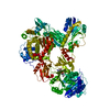
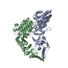
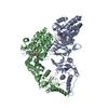
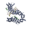
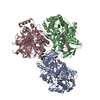
- PDB-6mfw: Crystal structure of a 4-domain construct of LgrA in the substrat... -
+ Open data

- Basic information
| Entry | Database: PDB / ID: 6mfw | |||||||||
|---|---|---|---|---|---|---|---|---|---|---|
| Title | Crystal structure of a 4-domain construct of LgrA in the substrate donation state | |||||||||
 Components Components | Linear gramicidin synthase subunit A | |||||||||
 Keywords Keywords |  LIGASE / nonribosomal peptide synthetase / tailoring domain / NRPS / enzyme / natural product / linear gramicidin LIGASE / nonribosomal peptide synthetase / tailoring domain / NRPS / enzyme / natural product / linear gramicidin | |||||||||
| Function / homology |  Function and homology information Function and homology informationamino acid activation for nonribosomal peptide biosynthetic process / secondary metabolite biosynthetic process / phosphopantetheine binding / ligase activity / antibiotic biosynthetic process / cytosolSimilarity search - Function | |||||||||
| Biological species |  Brevibacillus parabrevis (bacteria) Brevibacillus parabrevis (bacteria) | |||||||||
| Method | X-RAY DIFFRACTION / SYNCHROTRON / MOLECULAR REPLACEMENT / Resolution: 2.5 Å | |||||||||
 Authors Authors | Reimer, J.M. / Eivaskhani, M. / Schmeing, T.M. | |||||||||
| Funding support |  Canada, 2items Canada, 2items
| |||||||||
 Citation Citation | Journal: Science / Year: 2019 Title: Structures of a dimodular nonribosomal peptide synthetase reveal conformational flexibility. Authors: Reimer, J.M. / Eivaskhani, M. / Harb, I. / Guarne, A. / Weigt, M. / Schmeing, T.M. | |||||||||
| History |
|
- Structure visualization
| Structure viewer | Molecule: MolmilJmol/JSmol |
|---|
- Downloads & links
-Download
| PDBx/mmCIF format | 6mfw.cif.gz | 462.7 KB | Display | PDBx/mmCIF format |
|---|---|---|---|---|
| PDB format | pdb6mfw.ent.gz | 372.6 KB | Display | PDB format |
| PDBx/mmJSON format | 6mfw.json.gz | Tree view | PDBx/mmJSON format | |
| Others |  Other downloads Other downloads |
-Validation report
| Arichive directory | https://data.pdbj.org/pub/pdb/validation_reports/mf/6mfwftp://data.pdbj.org/pub/pdb/validation_reports/mf/6mfw | HTTPS FTP |
|---|
-Related structure data
| Related structure data |  6mfxC 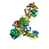 6mfyC 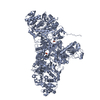 6mfzC 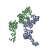 6mg0C 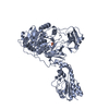 5es5S S: Starting model for refinement C: citing same article ( |
|---|---|
| Similar structure data |


-Links
 PDBj
PDBj


- Assembly
| Deposited unit | 
| ||||||||||
|---|---|---|---|---|---|---|---|---|---|---|---|
| 1 |
| ||||||||||
| Unit cell |
|
-Components
-Protein , 1 types, 1 molecules A
| #1: Protein | Mass: 138057.391 Da / Num. of mol.: 1 / Fragment: UNP residues 2-1198 Source method: isolated from a genetically manipulated source Source: (gene. exp.) Brevibacillus parabrevis (bacteria) / Gene: lgrA / Production host: Escherichia coli (E. coli) / References: UniProt: Q70LM7 |
|---|
-Non-polymers , 7 types, 185 molecules 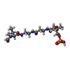


| #2: Chemical | ChemComp-JQG / ( Mass: 467.431 Da / Num. of mol.: 1 / Source method: obtained synthetically / Formula: C17H32N4O9P Mass: 467.431 Da / Num. of mol.: 1 / Source method: obtained synthetically / Formula: C17H32N4O9P |
|---|---|
| #3: Chemical | ChemComp-FON /  Mass: 473.439 Da / Num. of mol.: 1 / Source method: obtained synthetically / Formula: C20H23N7O7 Mass: 473.439 Da / Num. of mol.: 1 / Source method: obtained synthetically / Formula: C20H23N7O7 |
| #4: Chemical | ChemComp-APC /  Mass: 505.208 Da / Num. of mol.: 1 / Source method: obtained synthetically / Formula: C11H18N5O12P3 / Comment: AMP-CPP, energy-carrying molecule analogue*YM Mass: 505.208 Da / Num. of mol.: 1 / Source method: obtained synthetically / Formula: C11H18N5O12P3 / Comment: AMP-CPP, energy-carrying molecule analogue*YM |
| #5: Chemical | ChemComp-VAL / Valine Type: L-peptide linking / Mass: 117.146 Da / Num. of mol.: 1 / Source method: obtained synthetically / Formula: C5H11NO2 Type: L-peptide linking / Mass: 117.146 Da / Num. of mol.: 1 / Source method: obtained synthetically / Formula: C5H11NO2 |
| #6: Chemical | ChemComp-PO4 / Phosphate Mass: 94.971 Da / Num. of mol.: 1 / Source method: obtained synthetically / Formula: PO4 Mass: 94.971 Da / Num. of mol.: 1 / Source method: obtained synthetically / Formula: PO4 |
| #7: Chemical | ChemComp-MG /  Mass: 24.305 Da / Num. of mol.: 1 / Source method: obtained synthetically / Formula: Mg Mass: 24.305 Da / Num. of mol.: 1 / Source method: obtained synthetically / Formula: Mg |
| #8: Water | ChemComp-HOH / WaterMass: 18.015 Da / Num. of mol.: 179 / Source method: isolated from a natural source / Formula: H2O |
-Experimental details
-Experiment
| Experiment | Method: X-RAY DIFFRACTION / Number of used crystals: 1 |
|---|
- Sample preparation
| Crystal | Density Matthews: 2.68 Å3/Da / Density % sol: 54.02 % |
|---|---|
| Crystal grow | Temperature: 295 K / Method: vapor diffusion, sitting drop / pH: 7.7 Details: Protein + 2.5 mM AMPcPP + 2.5 mM valine + 2 mM N5-fTHF in 2.5 mM magnesium chloride, 15% PEG4000, 100 mM Tris, pH 7.78, 25 mM Tris, pH 7.5, 100 mM sodium chloride equilibrated against a ...Details: Protein + 2.5 mM AMPcPP + 2.5 mM valine + 2 mM N5-fTHF in 2.5 mM magnesium chloride, 15% PEG4000, 100 mM Tris, pH 7.78, 25 mM Tris, pH 7.5, 100 mM sodium chloride equilibrated against a reservoir containing 2.5 mM magnesium chloride, 18% PEG4000, 150 mM sodium chloride, 100 mM Tris, pH 7.7, drops streak-seeded from a seed stock of LgrA (F-A-PCP-C) crystals |
-Data collection
| Diffraction | Mean temperature: 100 K / Serial crystal experiment: N |
|---|---|
| Diffraction source | Source: SYNCHROTRON / Site: CLSI / Beamline: 08ID-1 / Wavelength: 0.979 Å |
| Detector | Type: DECTRIS PILATUS3 S 6M / Detector: PIXEL / Date: May 14, 2017 |
| Radiation | Monochromator: double crystal Si(111) / Protocol: SINGLE WAVELENGTH / Monochromatic (M) / Laue (L): M / Scattering type: x-ray |
| Radiation wavelength | Wavelength: 0.979 Å / Relative weight: 1 |
| Reflection | Resolution: 2.5→69.333 Å / Num. obs: 50890 / % possible obs: 100 % / Redundancy: 7.2 % / Biso Wilson estimate: 40.8 Å2 / Net I/σ(I): 10.1 |
| Reflection shell | Resolution: 2.5→2.58 Å / Num. unique obs: 4629 / % possible all: 99.9 |
- Processing
| Software |
| |||||||||||||||||||||||||||||||||||||||||||||||||||||||||||||||||||||||||||||||||||||||||||||||||||||||||
|---|---|---|---|---|---|---|---|---|---|---|---|---|---|---|---|---|---|---|---|---|---|---|---|---|---|---|---|---|---|---|---|---|---|---|---|---|---|---|---|---|---|---|---|---|---|---|---|---|---|---|---|---|---|---|---|---|---|---|---|---|---|---|---|---|---|---|---|---|---|---|---|---|---|---|---|---|---|---|---|---|---|---|---|---|---|---|---|---|---|---|---|---|---|---|---|---|---|---|---|---|---|---|---|---|---|---|
| Refinement | Method to determine structure: MOLECULAR REPLACEMENT Starting model: PDB entry 5ES5 Resolution: 2.5→69.333 Å / SU ML: 0.33 / Cross valid method: FREE R-VALUE / σ(F): 1.33 / Phase error: 26.31
| |||||||||||||||||||||||||||||||||||||||||||||||||||||||||||||||||||||||||||||||||||||||||||||||||||||||||
| Solvent computation | Shrinkage radii: 0.9 Å / VDW probe radii: 1.11 Å | |||||||||||||||||||||||||||||||||||||||||||||||||||||||||||||||||||||||||||||||||||||||||||||||||||||||||
| Refinement step | Cycle: LAST / Resolution: 2.5→69.333 Å
| |||||||||||||||||||||||||||||||||||||||||||||||||||||||||||||||||||||||||||||||||||||||||||||||||||||||||
| Refine LS restraints |
| |||||||||||||||||||||||||||||||||||||||||||||||||||||||||||||||||||||||||||||||||||||||||||||||||||||||||
| LS refinement shell |
|
