 Movie
Movie Controller

[English] 日本語
 Yorodumi
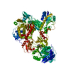
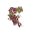
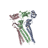
- PDB-5oqn: Crystal structure of the S. cerevisiae condensin Ycg1-Brn1 subcom... -
+ Open data

- Basic information
| Entry | Database: PDB / ID: 5oqn | |||||||||
|---|---|---|---|---|---|---|---|---|---|---|
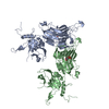
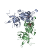
| Title | Crystal structure of the S. cerevisiae condensin Ycg1-Brn1 subcomplex bound to DNA (short kleisin loop) | |||||||||
 Components Components |
| |||||||||
 Keywords Keywords |  CELL CYCLE / kleisin / HEAT repeat / DNA-binding / SMC complex CELL CYCLE / kleisin / HEAT repeat / DNA-binding / SMC complex | |||||||||
| Function / homology |  Function and homology information Function and homology informationnegative regulation of meiotic DNA double-strand break formation / meiotic chromosome condensation / Condensation of Prometaphase Chromosomes / tRNA gene clustering / meiotic chromosome separation / condensin complex / rDNA chromatin condensation / synaptonemal complex assembly / mitotic chromosome condensation / chromosome condensation ...negative regulation of meiotic DNA double-strand break formation / meiotic chromosome condensation / Condensation of Prometaphase Chromosomes / tRNA gene clustering / meiotic chromosome separation / condensin complex / rDNA chromatin condensation / synaptonemal complex assembly / mitotic chromosome condensation / chromosome condensation / mitotic sister chromatid segregation / condensed chromosome / cell division / chromatin binding / nucleus / cytoplasmSimilarity search - Function | |||||||||
| Biological species |  Saccharomyces cerevisiae (brewer's yeast) Saccharomyces cerevisiae (brewer's yeast)synthetic construct (others) | |||||||||
| Method | X-RAY DIFFRACTION / SYNCHROTRON / MOLECULAR REPLACEMENT / Resolution: 3.15 Å | |||||||||
 Authors Authors | Kschonsak, M. / Hassler, M. / Haering, C.H. | |||||||||
| Funding support |  Germany, 2items Germany, 2items
| |||||||||
 Citation Citation | Journal: Cell / Year: 2017 Title: Structural Basis for a Safety-Belt Mechanism That Anchors Condensin to Chromosomes. Authors: Kschonsak, M. / Merkel, F. / Bisht, S. / Metz, J. / Rybin, V. / Hassler, M. / Haering, C.H. | |||||||||
| History |
|
- Structure visualization
| Structure viewer | Molecule: MolmilJmol/JSmol |
|---|
- Downloads & links
-Download
| PDBx/mmCIF format | 5oqn.cif.gz | 389.7 KB | Display | PDBx/mmCIF format |
|---|---|---|---|---|
| PDB format | pdb5oqn.ent.gz | 314.4 KB | Display | PDB format |
| PDBx/mmJSON format | 5oqn.json.gz | Tree view | PDBx/mmJSON format | |
| Others |  Other downloads Other downloads |
-Validation report
| Arichive directory | https://data.pdbj.org/pub/pdb/validation_reports/oq/5oqnftp://data.pdbj.org/pub/pdb/validation_reports/oq/5oqn | HTTPS FTP |
|---|
-Related structure data
| Related structure data | 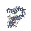 5oqoC  5oqpSC  5oqqC  5oqrC S: Starting model for refinement C: citing same article ( |
|---|---|
| Similar structure data |


-Links
 PDBj
PDBj


- Assembly
| Deposited unit | 
| ||||||||
|---|---|---|---|---|---|---|---|---|---|
| 1 |
| ||||||||
| Unit cell |
|
-Components
| #1: Protein | / CAPG homolog Mass: 99715.875 Da / Num. of mol.: 1 Source method: isolated from a genetically manipulated source Details: loop deletion: 499-555 Source: (gene. exp.) Saccharomyces cerevisiae (strain ATCC 204508 / S288c) (yeast)Gene: YCG1, YCS5, YDR325W / Plasmid: pETMCN / Production host:  Escherichia coli (E. coli) / Strain (production host): K12 / Variant (production host): Rosetta (DE3) pLysS / References: UniProt: Q06680 Escherichia coli (E. coli) / Strain (production host): K12 / Variant (production host): Rosetta (DE3) pLysS / References: UniProt: Q06680 |
|---|---|
| #2: Protein | / Barren homolog / CAPH homolog Mass: 15124.465 Da / Num. of mol.: 1 Source method: isolated from a genetically manipulated source Details: 384-529 loop deletion: 418-444 Source: (gene. exp.) Saccharomyces cerevisiae (strain ATCC 204508 / S288c) (yeast)Gene: BRN1, YBL097W, YBL0830 / Plasmid: pETMCN / Production host: Escherichia coli (E. coli) / Strain (production host): K12 / Variant (production host): Rosetta (DE3) pLysS / References: UniProt: P38170 |
| #3: DNA chain | Mass: 5515.590 Da / Num. of mol.: 2 / Source method: obtained synthetically / Source: (synth.) synthetic construct (others) |
-Experimental details
-Experiment
| Experiment | Method: X-RAY DIFFRACTION / Number of used crystals: 1 |
|---|
- Sample preparation
| Crystal | Density Matthews: 3.2 Å3/Da / Density % sol: 61.57 % |
|---|---|
| Crystal grow | Temperature: 280 K / Method: vapor diffusion Details: 4% (w/v) PEG 4000, 0.05 M Na citrate pH 5.6, 5 mM MgSO4 |
-Data collection
| Diffraction | Mean temperature: 100 K |
|---|---|
| Diffraction source | Source: SYNCHROTRON / Site: ESRF  / Beamline: ID29 / Wavelength: 0.979 Å / Beamline: ID29 / Wavelength: 0.979 Å |
| Detector | Type: DECTRIS PILATUS 6M / Detector: PIXEL / Date: Nov 17, 2016 |
| Radiation | Protocol: SINGLE WAVELENGTH / Monochromatic (M) / Laue (L): M / Scattering type: x-ray |
| Radiation wavelength | Wavelength: 0.979 Å / Relative weight: 1 |
| Reflection | Resolution: 3.15→48.68 Å / Num. obs: 28612 / % possible obs: 100 % / Redundancy: 12.3 % / CC1/2: 0.999 / Rmerge(I) obs: 0.198 / Rpim(I) all: 0.059 / Net I/σ(I): 8.4 |
| Reflection shell | Resolution: 3.15→3.32 Å / Redundancy: 8.6 % / Rmerge(I) obs: 1.115 / Mean I/σ(I) obs: 1.9 / Num. unique obs: 4129 / CC1/2: 0.287 / Rpim(I) all: 0.401 / % possible all: 100 |
- Processing
| Software |
| |||||||||||||||||||||||||||||||||||||||||||||||||||||||||||||||||||||||||||||||||||||||||||||||||||||||||
|---|---|---|---|---|---|---|---|---|---|---|---|---|---|---|---|---|---|---|---|---|---|---|---|---|---|---|---|---|---|---|---|---|---|---|---|---|---|---|---|---|---|---|---|---|---|---|---|---|---|---|---|---|---|---|---|---|---|---|---|---|---|---|---|---|---|---|---|---|---|---|---|---|---|---|---|---|---|---|---|---|---|---|---|---|---|---|---|---|---|---|---|---|---|---|---|---|---|---|---|---|---|---|---|---|---|---|
| Refinement | Method to determine structure: MOLECULAR REPLACEMENT Starting model: 5OQP Resolution: 3.15→47.343 Å / SU ML: 0.6 / Cross valid method: FREE R-VALUE / σ(F): 1.36 / Phase error: 33.46 / Stereochemistry target values: ML
| |||||||||||||||||||||||||||||||||||||||||||||||||||||||||||||||||||||||||||||||||||||||||||||||||||||||||
| Solvent computation | Shrinkage radii: 0.9 Å / VDW probe radii: 1.11 Å / Solvent model: FLAT BULK SOLVENT MODEL | |||||||||||||||||||||||||||||||||||||||||||||||||||||||||||||||||||||||||||||||||||||||||||||||||||||||||
| Refinement step | Cycle: LAST / Resolution: 3.15→47.343 Å
| |||||||||||||||||||||||||||||||||||||||||||||||||||||||||||||||||||||||||||||||||||||||||||||||||||||||||
| Refine LS restraints |
| |||||||||||||||||||||||||||||||||||||||||||||||||||||||||||||||||||||||||||||||||||||||||||||||||||||||||
| LS refinement shell |
|
