 Movie
Movie Controller

[English] 日本語
 Yorodumi
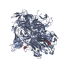
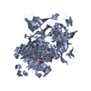
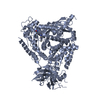
- PDB-4rgg: Structure of the lactococcal phage 1358 receptor binding protein ... -
+ Open data

- Basic information
| Entry | Database: PDB / ID: 4rgg | |||||||||
|---|---|---|---|---|---|---|---|---|---|---|
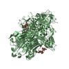
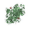
| Title | Structure of the lactococcal phage 1358 receptor binding protein in complex with GlcNAc-1P | |||||||||
 Components Components | Putative phage structural protein | |||||||||
 Keywords Keywords |  VIRAL PROTEIN / Alpha/beta protein / phage receptor bonding protein (anti-receptor) / L. lactis SMQ-388 surface polysaccharides (pellicle) VIRAL PROTEIN / Alpha/beta protein / phage receptor bonding protein (anti-receptor) / L. lactis SMQ-388 surface polysaccharides (pellicle) | |||||||||
| Function / homology | Viral head domain of polysaccharide receptor-binding protein-like / Triple-stranded beta-helix / Phage fibre proteins / Jelly Rolls / Sandwich / Mainly Beta / metal ion binding / Chem-GN1 / Putative phage structural protein Function and homology information Function and homology information | |||||||||
| Biological species |  Lactococcus phage 1358 (virus) Lactococcus phage 1358 (virus) | |||||||||
| Method | X-RAY DIFFRACTION / MIR / Resolution: 2.15 Å | |||||||||
 Authors Authors | Spinelli, S. / Moineau, S. / Cambillau, C. | |||||||||
 Citation Citation | Journal: To be Published Title: Structure of the 1358 lactococcal phage RBP in complex with GlcNAc-1P Authors: Spinelli, S. / Moineau, S. / Cambillau, C. | |||||||||
| History |
|
- Structure visualization
| Structure viewer | Molecule: MolmilJmol/JSmol |
|---|
- Downloads & links
-Download
| PDBx/mmCIF format | 4rgg.cif.gz | 322.1 KB | Display | PDBx/mmCIF format |
|---|---|---|---|---|
| PDB format | pdb4rgg.ent.gz | 263.3 KB | Display | PDB format |
| PDBx/mmJSON format | 4rgg.json.gz | Tree view | PDBx/mmJSON format | |
| Others |  Other downloads Other downloads |
-Validation report
| Arichive directory | https://data.pdbj.org/pub/pdb/validation_reports/rg/4rggftp://data.pdbj.org/pub/pdb/validation_reports/rg/4rgg | HTTPS FTP |
|---|
-Related structure data
| Related structure data |  4l92S S: Starting model for refinement |
|---|---|
| Similar structure data |


-Links
 PDBj
PDBj- Assembly
| Deposited unit | 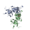
| |||||||||||||||||||||
|---|---|---|---|---|---|---|---|---|---|---|---|---|---|---|---|---|---|---|---|---|---|---|
| 1 | 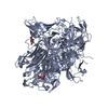
| |||||||||||||||||||||
| 2 | 
| |||||||||||||||||||||
| Unit cell |
| |||||||||||||||||||||
| Components on special symmetry positions |
| |||||||||||||||||||||
| Noncrystallographic symmetry (NCS) | NCS domain:
NCS domain segments: Component-ID: 0 / Ens-ID: 1 / Beg auth comp-ID: VAL / Beg label comp-ID: VAL / End auth comp-ID: PHE / End label comp-ID: PHE / Refine code: 0 / Auth seq-ID: 2 - 393 / Label seq-ID: 2 - 393
|
-Components
| #1: Protein | Mass: 43342.027 Da / Num. of mol.: 2 / Fragment: UNP residues 2-393 Source method: isolated from a genetically manipulated source Source: (gene. exp.) Lactococcus phage 1358 (virus) / Gene: ORF20 / Production host:  Escherichia coli (E. coli) / Strain (production host): BL21 / References: UniProt: D3W0F1 Escherichia coli (E. coli) / Strain (production host): BL21 / References: UniProt: D3W0F1#2: Sugar |   Type: D-saccharide / Mass: 301.188 Da / Num. of mol.: 2 / Source method: obtained synthetically / Formula: C8H16NO9P Type: D-saccharide / Mass: 301.188 Da / Num. of mol.: 2 / Source method: obtained synthetically / Formula: C8H16NO9P#3: Water | ChemComp-HOH / | Water Mass: 18.015 Da / Num. of mol.: 502 / Source method: isolated from a natural source / Formula: H2O Mass: 18.015 Da / Num. of mol.: 502 / Source method: isolated from a natural source / Formula: H2O |
|---|
-Experimental details
-Experiment
| Experiment | Method: X-RAY DIFFRACTION / Number of used crystals: 1 |
|---|
- Sample preparation
| Crystal | Density Matthews: 4.39 Å3/Da / Density % sol: 71.98 % |
|---|---|
| Crystal grow | Temperature: 293 K / pH: 6.2 Details: 20% w/v polyethyleneglycol 1000, 100 mM sodium/potassium phosphate buffer, pH 6.2, 200 mM NaCl, VAPOR DIFFUSION, SITTING DROP, temperature 293K |
-Data collection
| Diffraction | Mean temperature: 100 K |
|---|---|
| Diffraction source | Source: ROTATING ANODE / Type: BRUKER AXS MICROSTAR / Wavelength: 1.5418 |
| Detector | Type: DECTRIS PILATUS 6M / Detector: PIXEL / Date: May 3, 2014 |
| Radiation | Monochromator: MIRRORS / Protocol: SINGLE WAVELENGTH / Monochromatic (M) / Laue (L): M / Scattering type: x-ray |
| Radiation wavelength | Wavelength: 1.5418 Å / Relative weight: 1 |
| Reflection | Resolution: 2.15→48 Å / Num. obs: 81936 / % possible obs: 99.9 % / Observed criterion σ(I): 0 / Redundancy: 12.3 % / Biso Wilson estimate: 49 Å2 / Rmerge(I) obs: 0.089 / Net I/σ(I): 20.5 |
| Reflection shell | Resolution: 2.15→2.21 Å / Rmerge(I) obs: 0.81 / Mean I/σ(I) obs: 2.43 / % possible all: 99.9 |
- Processing
| Software |
| ||||||||||||||||||||||||||||||||||||||||||||||||||||||||||||||||||||||||||||||||||||||||||||||||||||||||||||||||||||||||||||||||||||||||||||||||||||||||||||||||||||||||||||||||||||||
|---|---|---|---|---|---|---|---|---|---|---|---|---|---|---|---|---|---|---|---|---|---|---|---|---|---|---|---|---|---|---|---|---|---|---|---|---|---|---|---|---|---|---|---|---|---|---|---|---|---|---|---|---|---|---|---|---|---|---|---|---|---|---|---|---|---|---|---|---|---|---|---|---|---|---|---|---|---|---|---|---|---|---|---|---|---|---|---|---|---|---|---|---|---|---|---|---|---|---|---|---|---|---|---|---|---|---|---|---|---|---|---|---|---|---|---|---|---|---|---|---|---|---|---|---|---|---|---|---|---|---|---|---|---|---|---|---|---|---|---|---|---|---|---|---|---|---|---|---|---|---|---|---|---|---|---|---|---|---|---|---|---|---|---|---|---|---|---|---|---|---|---|---|---|---|---|---|---|---|---|---|---|---|---|
| Refinement | Method to determine structure: MIR Starting model: PDB ENTRY 4L92 Resolution: 2.15→47.89 Å / Cor.coef. Fo:Fc: 0.957 / Cor.coef. Fo:Fc free: 0.95 / SU B: 7.136 / SU ML: 0.096 / Cross valid method: THROUGHOUT / σ(F): 0 / ESU R: 0.14 / ESU R Free: 0.129 / Stereochemistry target values: MAXIMUM LIKELIHOOD / Details: HYDROGENS HAVE BEEN ADDED IN THE RIDING POSITIONS
| ||||||||||||||||||||||||||||||||||||||||||||||||||||||||||||||||||||||||||||||||||||||||||||||||||||||||||||||||||||||||||||||||||||||||||||||||||||||||||||||||||||||||||||||||||||||
| Solvent computation | Ion probe radii: 0.8 Å / Shrinkage radii: 0.8 Å / VDW probe radii: 1.2 Å / Solvent model: MASK | ||||||||||||||||||||||||||||||||||||||||||||||||||||||||||||||||||||||||||||||||||||||||||||||||||||||||||||||||||||||||||||||||||||||||||||||||||||||||||||||||||||||||||||||||||||||
| Displacement parameters | Biso mean: 45.25 Å2
| ||||||||||||||||||||||||||||||||||||||||||||||||||||||||||||||||||||||||||||||||||||||||||||||||||||||||||||||||||||||||||||||||||||||||||||||||||||||||||||||||||||||||||||||||||||||
| Refine analyze | Luzzati coordinate error free: 0.322 Å | ||||||||||||||||||||||||||||||||||||||||||||||||||||||||||||||||||||||||||||||||||||||||||||||||||||||||||||||||||||||||||||||||||||||||||||||||||||||||||||||||||||||||||||||||||||||
| Refinement step | Cycle: LAST / Resolution: 2.15→47.89 Å
| ||||||||||||||||||||||||||||||||||||||||||||||||||||||||||||||||||||||||||||||||||||||||||||||||||||||||||||||||||||||||||||||||||||||||||||||||||||||||||||||||||||||||||||||||||||||
| Refine LS restraints |
|