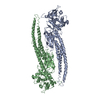
Entry Database : PDB / ID : 6mbwTitle Structure of Transcription Factor Signal transducer and activator of transcription 5B Keywords / Function / homology Function Domain/homology Component
/ / / / / / / / / / / / / / / / / / / / / / / / / / / / / / / / / / / / / / / / / / / / / / / / / / / / / / / / / / / / / / / / / / / / / / / / / / / / / / / / / / / / / / / / / / / / / / / / / / / / / / / / / / / / / / / / / / / / / / / / / / / Biological species Homo sapiens (human)Method / / / / Resolution : 3.29 Å Authors Seo, H.-S. / Dhe-Paganon, S. Funding support Organization Grant number Country Canadian Institutes of Health Research (CIHR)
Journal : Nat Commun / Year : 2019Title : Structural and functional consequences of the STAT5BN642H driver mutation.Authors: de Araujo, E.D. / Erdogan, F. / Neubauer, H.A. / Meneksedag-Erol, D. / Manaswiyoungkul, P. / Eram, M.S. / Seo, H.S. / Qadree, A.K. / Israelian, J. / Orlova, A. / Suske, T. / Pham, H.T.T. / ... Authors : de Araujo, E.D. / Erdogan, F. / Neubauer, H.A. / Meneksedag-Erol, D. / Manaswiyoungkul, P. / Eram, M.S. / Seo, H.S. / Qadree, A.K. / Israelian, J. / Orlova, A. / Suske, T. / Pham, H.T.T. / Boersma, A. / Tangermann, S. / Kenner, L. / Rulicke, T. / Dong, A. / Ravichandran, M. / Brown, P.J. / Audette, G.F. / Rauscher, S. / Dhe-Paganon, S. / Moriggl, R. / Gunning, P.T. History Deposition Aug 30, 2018 Deposition site / Processing site Revision 1.0 Jun 19, 2019 Provider / Type Revision 1.1 Jan 8, 2020 Group / Category / Item Revision 1.2 Oct 11, 2023 Group / Database references / Refinement descriptionCategory chem_comp_atom / chem_comp_bond ... chem_comp_atom / chem_comp_bond / database_2 / pdbx_initial_refinement_model Item / _database_2.pdbx_database_accession
Show all Show less
 Movie
Movie Controller


 Open data
 Basic information
 Components
 Keywords
 TRANSCRIPTION ACTIVATOR /
  Function and homology information
 Authors
 Canada, 1items
  Citation
 Structure visualization
 Downloads & links
 Other downloads


 PDBj
PDBj


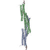
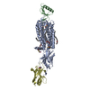
 Assembly

 Mass: 18.015 Da / Num. of mol.: 3 / Source method: isolated from a natural source / Formula: H2O
 Sample preparation
 / Beamline: 24-ID-E / Wavelength: 0.9792 Å
 Processing