 Movie
Movie Controller


+ Open data

- Basic information
| Entry | Database: PDB / ID: 6m9t | ||||||
|---|---|---|---|---|---|---|---|
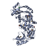
| Title | Crystal structure of EP3 receptor bound to misoprostol-FA | ||||||
 Components Components | Prostaglandin E2 receptor EP3 subtype, Endolysin chimera | ||||||
 Keywords Keywords |  MEMBRANE PROTEIN / GPCR / prostaglandin E2 receptor 3 (EP3) / prostaglandin analogue / misoprostol-FA (biologically active free acid) / XFEL / LCP / T4L MEMBRANE PROTEIN / GPCR / prostaglandin E2 receptor 3 (EP3) / prostaglandin analogue / misoprostol-FA (biologically active free acid) / XFEL / LCP / T4L | ||||||
| Function / homology |  Function and homology information Function and homology informationnegative regulation of gastric acid secretion / intestine smooth muscle contraction / prostaglandin E receptor activity / Prostanoid ligand receptors / cell death / positive regulation of fever generation / viral release from host cell by cytolysis / peptidoglycan catabolic process / adenylate cyclase-activating G protein-coupled receptor signaling pathway / cell wall macromolecule catabolic process ...negative regulation of gastric acid secretion / intestine smooth muscle contraction / prostaglandin E receptor activity / Prostanoid ligand receptors / cell death / positive regulation of fever generation / viral release from host cell by cytolysis / peptidoglycan catabolic process / adenylate cyclase-activating G protein-coupled receptor signaling pathway / cell wall macromolecule catabolic process / lysozyme / nuclear envelope / phospholipase C-activating G protein-coupled receptor signaling pathway / lysozyme activity / G alpha (i) signalling events / positive regulation of cytosolic calcium ion concentration / host cell cytoplasm / defense response to bacterium / inflammatory response / G protein-coupled receptor signaling pathway / membrane / plasma membraneSimilarity search - Function | ||||||
| Biological species |  Homo sapiens (human) Homo sapiens (human) Enterobacteria phage T4 (virus) Enterobacteria phage T4 (virus) | ||||||
| Method | X-RAY DIFFRACTION / FREE ELECTRON LASER / MOLECULAR REPLACEMENT / Resolution: 2.5 Å | ||||||
 Authors Authors | Audet, M. / White, K.L. / Breton, B. / Zarzycka, B. / Han, G.W. / Lu, Y. / Gati, C. / Batyuk, A. / Popov, P. / Velasquez, J. ...Audet, M. / White, K.L. / Breton, B. / Zarzycka, B. / Han, G.W. / Lu, Y. / Gati, C. / Batyuk, A. / Popov, P. / Velasquez, J. / Manahan, D. / Hu, H. / Weierstall, U. / Liu, W. / Shui, W. / Katrich, V. / Cherezov, V. / Hanson, M.A. / Stevens, R.C. | ||||||
 Citation Citation | Journal: Nat. Chem. Biol. / Year: 2019 Title: Crystal structure of misoprostol bound to the labor inducer prostaglandin E2receptor. Authors: Audet, M. / White, K.L. / Breton, B. / Zarzycka, B. / Han, G.W. / Lu, Y. / Gati, C. / Batyuk, A. / Popov, P. / Velasquez, J. / Manahan, D. / Hu, H. / Weierstall, U. / Liu, W. / Shui, W. / ...Authors: Audet, M. / White, K.L. / Breton, B. / Zarzycka, B. / Han, G.W. / Lu, Y. / Gati, C. / Batyuk, A. / Popov, P. / Velasquez, J. / Manahan, D. / Hu, H. / Weierstall, U. / Liu, W. / Shui, W. / Katritch, V. / Cherezov, V. / Hanson, M.A. / Stevens, R.C. | ||||||
| History |
|
- Structure visualization
| Structure viewer | Molecule: MolmilJmol/JSmol |
|---|
- Downloads & links
-Download
| PDBx/mmCIF format | 6m9t.cif.gz | 201.5 KB | Display | PDBx/mmCIF format |
|---|---|---|---|---|
| PDB format | pdb6m9t.ent.gz | 158.6 KB | Display | PDB format |
| PDBx/mmJSON format | 6m9t.json.gz | Tree view | PDBx/mmJSON format | |
| Others |  Other downloads Other downloads |
-Validation report
| Arichive directory | https://data.pdbj.org/pub/pdb/validation_reports/m9/6m9tftp://data.pdbj.org/pub/pdb/validation_reports/m9/6m9t | HTTPS FTP |
|---|
-Related structure data


-Links
 PDBj
PDBj


- Assembly
| Deposited unit | 
| ||||||||
|---|---|---|---|---|---|---|---|---|---|
| 1 |
| ||||||||
| Unit cell |
| ||||||||
| Details | Authors state that the biological unit is unknown. |
-Components
-Protein , 1 types, 1 molecules A
| #1: Protein | Mass: 60023.445 Da / Num. of mol.: 1 Fragment: EP3 UNP residues 2-259,273-353 with intervening lysozyme Source method: isolated from a genetically manipulated source Source: (gene. exp.) Homo sapiens (human), (gene. exp.) Enterobacteria phage T4 (virus)Gene: PTGER3 / Production host:   Spodoptera frugiperda (fall armyworm) / References: UniProt: P43115, UniProt: P00720, lysozyme Spodoptera frugiperda (fall armyworm) / References: UniProt: P43115, UniProt: P00720, lysozyme |
|---|
-Non-polymers , 5 types, 15 molecules 


| #2: Chemical | ChemComp-J9P / ( Mass: 368.508 Da / Num. of mol.: 1 / Source method: obtained synthetically / Formula: C21H36O5 Mass: 368.508 Da / Num. of mol.: 1 / Source method: obtained synthetically / Formula: C21H36O5 | ||||||
|---|---|---|---|---|---|---|---|
| #3: Chemical | ChemComp-SO4 / Sulfate Mass: 96.063 Da / Num. of mol.: 4 / Source method: obtained synthetically / Formula: SO4 Mass: 96.063 Da / Num. of mol.: 4 / Source method: obtained synthetically / Formula: SO4#4: Chemical | ChemComp-OLC / ( |  Mass: 356.540 Da / Num. of mol.: 1 / Source method: obtained synthetically / Formula: C21H40O4 Mass: 356.540 Da / Num. of mol.: 1 / Source method: obtained synthetically / Formula: C21H40O4#5: Chemical | ChemComp-OLA / Oleic acid Mass: 282.461 Da / Num. of mol.: 4 / Source method: obtained synthetically / Formula: C18H34O2 Mass: 282.461 Da / Num. of mol.: 4 / Source method: obtained synthetically / Formula: C18H34O2#6: Water | ChemComp-HOH / | WaterMass: 18.015 Da / Num. of mol.: 5 / Source method: isolated from a natural source / Formula: H2O |
-Experimental details
-Experiment
| Experiment | Method: X-RAY DIFFRACTION / Number of used crystals: 1 |
|---|
- Sample preparation
| Crystal | Density Matthews: 2.66 Å3/Da / Density % sol: 53.72 % |
|---|---|
| Crystal grow | Temperature: 293 K / Method: lipidic cubic phase Details: 100 mM sodium citrate, pH 3.8-4.2, 10-35 mM magnesium sulfate, 20-23% v/v PEG400, 2.5% Jeffamine M-600 |
-Data collection
| Diffraction | Mean temperature: 293 K / Serial crystal experiment: Y |
|---|---|
| Diffraction source | Source: FREE ELECTRON LASER / Site: SLAC LCLS  / Beamline: CXI / Wavelength: 1.302 Å / Beamline: CXI / Wavelength: 1.302 Å |
| Detector | Type: CS-PAD CXI-1 / Detector: PIXEL / Date: Aug 19, 2017 |
| Radiation | Protocol: SINGLE WAVELENGTH / Monochromatic (M) / Laue (L): M / Scattering type: x-ray |
| Radiation wavelength | Wavelength: 1.302 Å / Relative weight: 1 |
| Reflection | Resolution: 2.5→30.03 Å / Num. obs: 22151 / % possible obs: 100 % / Redundancy: 223.9 % / Biso Wilson estimate: 74.95 Å2 / CC1/2: 0.9944 / R split: 0.01 / Net I/σ(I): 4.2 |
| Reflection shell | Resolution: 2.5→2.62 Å / Redundancy: 90.5 % / Mean I/σ(I) obs: 0.25 / Num. unique obs: 22097 / CC1/2: 0.193 / R split: 4.791 / % possible all: 100 |
| Serial crystallography measurement | Collection time total: 1.75 hours / Collimation: KB mirrors / Pulse duration: 40 fsec. / Pulse photon energy: 9.52 keV / XFEL pulse repetition rate: 120 Hz |
| Serial crystallography sample delivery | Method: injection |
| Serial crystallography sample delivery injection | Carrier solvent: LCP / Flow rate: 0.22 µL/min / Injector diameter: 50 µm / Injector nozzle: LCP / Power by: HPLC pump |
| Serial crystallography data reduction | Crystal hits: 32143 / Frames failed index: 3643 / Frames indexed: 28500 / Frames total: 1311952 / Lattices indexed: 28500 / XFEL run numbers: 146-156 |
- Processing
| Software |
| ||||||||||||||||||||||||||||||||||||||||||||||||||||||||||||||||||||||||||||||||||||||||||||||||||||||||||||||||||
|---|---|---|---|---|---|---|---|---|---|---|---|---|---|---|---|---|---|---|---|---|---|---|---|---|---|---|---|---|---|---|---|---|---|---|---|---|---|---|---|---|---|---|---|---|---|---|---|---|---|---|---|---|---|---|---|---|---|---|---|---|---|---|---|---|---|---|---|---|---|---|---|---|---|---|---|---|---|---|---|---|---|---|---|---|---|---|---|---|---|---|---|---|---|---|---|---|---|---|---|---|---|---|---|---|---|---|---|---|---|---|---|---|---|---|---|
| Refinement | Method to determine structure: MOLECULAR REPLACEMENT Starting model: PDB entries 3P0G & 4EIY Resolution: 2.5→27.27 Å / Cor.coef. Fo:Fc: 0.956 / Cor.coef. Fo:Fc free: 0.95 / Rfactor Rfree error: 0 / SU R Cruickshank DPI: 0.337 / Cross valid method: THROUGHOUT / σ(F): 0 / SU R Blow DPI: 0.315 / SU Rfree Blow DPI: 0.234 / SU Rfree Cruickshank DPI: 0.242
| ||||||||||||||||||||||||||||||||||||||||||||||||||||||||||||||||||||||||||||||||||||||||||||||||||||||||||||||||||
| Displacement parameters | Biso mean: 128.85 Å2
| ||||||||||||||||||||||||||||||||||||||||||||||||||||||||||||||||||||||||||||||||||||||||||||||||||||||||||||||||||
| Refine analyze | Luzzati coordinate error obs: 0.56 Å | ||||||||||||||||||||||||||||||||||||||||||||||||||||||||||||||||||||||||||||||||||||||||||||||||||||||||||||||||||
| Refinement step | Cycle: 1 / Resolution: 2.5→27.27 Å
| ||||||||||||||||||||||||||||||||||||||||||||||||||||||||||||||||||||||||||||||||||||||||||||||||||||||||||||||||||
| Refine LS restraints |
| ||||||||||||||||||||||||||||||||||||||||||||||||||||||||||||||||||||||||||||||||||||||||||||||||||||||||||||||||||
| LS refinement shell | Resolution: 2.5→2.62 Å / Rfactor Rfree error: 0 / Total num. of bins used: 11
| ||||||||||||||||||||||||||||||||||||||||||||||||||||||||||||||||||||||||||||||||||||||||||||||||||||||||||||||||||
| Refinement TLS params. | Method: refined / Refine-ID: X-RAY DIFFRACTION
| ||||||||||||||||||||||||||||||||||||||||||||||||||||||||||||||||||||||||||||||||||||||||||||||||||||||||||||||||||
| Refinement TLS group |
|
