 Movie
Movie Controller


+ Open data

- Basic information
| Entry | Database: PDB / ID: 6m81 | ||||||
|---|---|---|---|---|---|---|---|
| Title | Crystal structure of TylM1 Y14F bound to SAH and dTDP-phenol | ||||||
 Components Components | dTDP-3-amino-3,6-dideoxy-alpha-D-glucopyranose N,N-dimethyltransferase | ||||||
 Keywords Keywords | TRANSFERASE / TylM1 / N-methyltransferase | ||||||
| Function / homology |  Function and homology informationdTDP-3-amino-3,6-dideoxy-alpha-D-glucopyranose N,N-dimethyltransferase / S-adenosylmethionine-dependent methyltransferase activity / antibiotic biosynthetic process / methylation / protein homodimerization activity Function and homology informationdTDP-3-amino-3,6-dideoxy-alpha-D-glucopyranose N,N-dimethyltransferase / S-adenosylmethionine-dependent methyltransferase activity / antibiotic biosynthetic process / methylation / protein homodimerization activitySimilarity search - Function | ||||||
| Biological species |  Streptomyces fradiae (bacteria) Streptomyces fradiae (bacteria) | ||||||
| Method | X-RAY DIFFRACTION / SYNCHROTRON / MOLECULAR REPLACEMENT / molecular replacement / Resolution: 1.782 Å | ||||||
 Authors Authors | Fick, R.J. / McDole, B.G. / Trievel, R.C. | ||||||
| Funding support |  United States, 1items United States, 1items
| ||||||
 Citation Citation | Journal: Biochemistry / Year: 2019 Title: Structural and Functional Characterization of Sulfonium Carbon-Oxygen Hydrogen Bonding in the Deoxyamino Sugar Methyltransferase TylM1. Authors: Fick, R.J. / Horowitz, S. / McDole, B.G. / Clay, M.C. / Mehl, R.A. / Al-Hashimi, H.M. / Scheiner, S. / Trievel, R.C. | ||||||
| History |
|
- Structure visualization
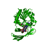
| Structure viewer | Molecule: MolmilJmol/JSmol |
|---|
- Downloads & links
-Download
| PDBx/mmCIF format | 6m81.cif.gz | 216.5 KB | Display | PDBx/mmCIF format |
|---|---|---|---|---|
| PDB format | pdb6m81.ent.gz | 171 KB | Display | PDB format |
| PDBx/mmJSON format | 6m81.json.gz | Tree view | PDBx/mmJSON format | |
| Others |  Other downloads Other downloads |
-Validation report
| Arichive directory | https://data.pdbj.org/pub/pdb/validation_reports/m8/6m81ftp://data.pdbj.org/pub/pdb/validation_reports/m8/6m81 | HTTPS FTP |
|---|
-Related structure data


-Links
 PDBj
PDBj- Assembly
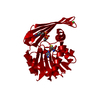
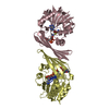
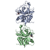
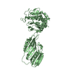
| Deposited unit | 
| ||||||||
|---|---|---|---|---|---|---|---|---|---|
| 1 | 
| ||||||||
| 2 | 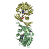
| ||||||||
| Unit cell |
|
-Components
| #1: Protein | / Tylosin biosynthesis protein M1 Mass: 28521.881 Da / Num. of mol.: 4 / Mutation: Y14F Source method: isolated from a genetically manipulated source Source: (gene. exp.) Streptomyces fradiae (bacteria) / Gene: tylM1, tylMI / Plasmid: pET31 / Production host: Escherichia coli (E. coli) / Strain (production host): Rosetta2(DE3)References: UniProt: P95748, dTDP-3-amino-3,6-dideoxy-alpha-D-glucopyranose N,N-dimethyltransferase#2: Chemical | ChemComp-SAH / S-Adenosyl-L-homocysteine  Type: L-peptide linking / Mass: 384.411 Da / Num. of mol.: 4 / Source method: obtained synthetically / Formula: C14H20N6O5S Type: L-peptide linking / Mass: 384.411 Da / Num. of mol.: 4 / Source method: obtained synthetically / Formula: C14H20N6O5S#3: Chemical | ChemComp-TLO /   Mass: 478.284 Da / Num. of mol.: 4 / Source method: obtained synthetically / Formula: C16H20N2O11P2 Mass: 478.284 Da / Num. of mol.: 4 / Source method: obtained synthetically / Formula: C16H20N2O11P2#4: Water | ChemComp-HOH / | Water Mass: 18.015 Da / Num. of mol.: 785 / Source method: isolated from a natural source / Formula: H2O Mass: 18.015 Da / Num. of mol.: 785 / Source method: isolated from a natural source / Formula: H2O |
|---|
-Experimental details
-Experiment
| Experiment | Method: X-RAY DIFFRACTION / Number of used crystals: 1 |
|---|
- Sample preparation
| Crystal | Density Matthews: 2.31 Å3/Da / Density % sol: 46.78 % / Mosaicity: 0.487 ° |
|---|---|
| Crystal grow | Temperature: 298 K / Method: vapor diffusion, hanging drop / pH: 7.46 Details: Precipitant:27% w/v PEG 3350, 20 mM sodium malonate, 1.25% v/v 2-propanol, 5.6 mM trimethylamine, 100 mM Hepes pH 7.46 Protein:20 mM sodium malonate pH 7.0, 100 mM sodium chloride, 5 mM ...Details: Precipitant:27% w/v PEG 3350, 20 mM sodium malonate, 1.25% v/v 2-propanol, 5.6 mM trimethylamine, 100 mM Hepes pH 7.46 Protein:20 mM sodium malonate pH 7.0, 100 mM sodium chloride, 5 mM AdoMet, 5 mM dTDP-phenol, 9 mg/mL TylM1 Y14F Protein:Precipitant 4:2uL |
-Data collection
| Diffraction | Mean temperature: 100 K | ||||||||||||||||||||||||||||||||||||||||||||||||||||||||||||||||||||||||||||||||||||||||||||||||||||||||||||||||||||||||||||||||||||||||||||||||
|---|---|---|---|---|---|---|---|---|---|---|---|---|---|---|---|---|---|---|---|---|---|---|---|---|---|---|---|---|---|---|---|---|---|---|---|---|---|---|---|---|---|---|---|---|---|---|---|---|---|---|---|---|---|---|---|---|---|---|---|---|---|---|---|---|---|---|---|---|---|---|---|---|---|---|---|---|---|---|---|---|---|---|---|---|---|---|---|---|---|---|---|---|---|---|---|---|---|---|---|---|---|---|---|---|---|---|---|---|---|---|---|---|---|---|---|---|---|---|---|---|---|---|---|---|---|---|---|---|---|---|---|---|---|---|---|---|---|---|---|---|---|---|---|---|---|
| Diffraction source | Source: SYNCHROTRON / Site: APS / Beamline: 21-ID-G / Wavelength: 0.9786 Å | ||||||||||||||||||||||||||||||||||||||||||||||||||||||||||||||||||||||||||||||||||||||||||||||||||||||||||||||||||||||||||||||||||||||||||||||||
| Detector | Type: MARMOSAIC 300 mm CCD / Detector: CCD / Date: Apr 8, 2014 | ||||||||||||||||||||||||||||||||||||||||||||||||||||||||||||||||||||||||||||||||||||||||||||||||||||||||||||||||||||||||||||||||||||||||||||||||
| Radiation | Protocol: SINGLE WAVELENGTH / Monochromatic (M) / Laue (L): M / Scattering type: x-ray | ||||||||||||||||||||||||||||||||||||||||||||||||||||||||||||||||||||||||||||||||||||||||||||||||||||||||||||||||||||||||||||||||||||||||||||||||
| Radiation wavelength | Wavelength: 0.9786 Å / Relative weight: 1 | ||||||||||||||||||||||||||||||||||||||||||||||||||||||||||||||||||||||||||||||||||||||||||||||||||||||||||||||||||||||||||||||||||||||||||||||||
| Reflection | Resolution: 1.78→50 Å / Num. obs: 96413 / % possible obs: 97.5 % / Redundancy: 7 % / Biso Wilson estimate: 22.61 Å2 / Rmerge(I) obs: 0.064 / Rpim(I) all: 0.026 / Rrim(I) all: 0.069 / Χ2: 2.238 / Net I/σ(I): 14.9 / Num. measured all: 678249 | ||||||||||||||||||||||||||||||||||||||||||||||||||||||||||||||||||||||||||||||||||||||||||||||||||||||||||||||||||||||||||||||||||||||||||||||||
| Reflection shell | Diffraction-ID: 1
|
-Phasing
| Phasing | Method: molecular replacement |
|---|
- Processing
| Software |
| |||||||||||||||||||||||||||||||||||||||||||||||||||||||||||||||||||||||||||||||||||||||||||||||||||||||||||||||||||||||||||||||||||||||||||||||||||||||||||||||||||||||||||||||||||||||||||||||||||||||||||||||||||||||||
|---|---|---|---|---|---|---|---|---|---|---|---|---|---|---|---|---|---|---|---|---|---|---|---|---|---|---|---|---|---|---|---|---|---|---|---|---|---|---|---|---|---|---|---|---|---|---|---|---|---|---|---|---|---|---|---|---|---|---|---|---|---|---|---|---|---|---|---|---|---|---|---|---|---|---|---|---|---|---|---|---|---|---|---|---|---|---|---|---|---|---|---|---|---|---|---|---|---|---|---|---|---|---|---|---|---|---|---|---|---|---|---|---|---|---|---|---|---|---|---|---|---|---|---|---|---|---|---|---|---|---|---|---|---|---|---|---|---|---|---|---|---|---|---|---|---|---|---|---|---|---|---|---|---|---|---|---|---|---|---|---|---|---|---|---|---|---|---|---|---|---|---|---|---|---|---|---|---|---|---|---|---|---|---|---|---|---|---|---|---|---|---|---|---|---|---|---|---|---|---|---|---|---|---|---|---|---|---|---|---|---|---|---|---|---|---|---|---|---|
| Refinement | Method to determine structure: MOLECULAR REPLACEMENT / Resolution: 1.782→36.956 Å / SU ML: 0.19 / Cross valid method: THROUGHOUT / σ(F): 1.34 / Phase error: 23.76 / Stereochemistry target values: ML
| |||||||||||||||||||||||||||||||||||||||||||||||||||||||||||||||||||||||||||||||||||||||||||||||||||||||||||||||||||||||||||||||||||||||||||||||||||||||||||||||||||||||||||||||||||||||||||||||||||||||||||||||||||||||||
| Solvent computation | Shrinkage radii: 0.9 Å / VDW probe radii: 1.11 Å / Solvent model: FLAT BULK SOLVENT MODEL | |||||||||||||||||||||||||||||||||||||||||||||||||||||||||||||||||||||||||||||||||||||||||||||||||||||||||||||||||||||||||||||||||||||||||||||||||||||||||||||||||||||||||||||||||||||||||||||||||||||||||||||||||||||||||
| Displacement parameters | Biso max: 69.21 Å2 / Biso mean: 27.962 Å2 / Biso min: 12.85 Å2 | |||||||||||||||||||||||||||||||||||||||||||||||||||||||||||||||||||||||||||||||||||||||||||||||||||||||||||||||||||||||||||||||||||||||||||||||||||||||||||||||||||||||||||||||||||||||||||||||||||||||||||||||||||||||||
| Refinement step | Cycle: final / Resolution: 1.782→36.956 Å
| |||||||||||||||||||||||||||||||||||||||||||||||||||||||||||||||||||||||||||||||||||||||||||||||||||||||||||||||||||||||||||||||||||||||||||||||||||||||||||||||||||||||||||||||||||||||||||||||||||||||||||||||||||||||||
| LS refinement shell | Refine-ID: X-RAY DIFFRACTION / Rfactor Rfree error: 0 / Total num. of bins used: 30
|
