 Movie
Movie Controller

[English] 日本語
 Yorodumi
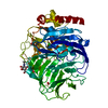
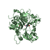
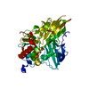
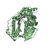
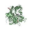
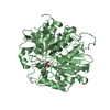
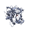
- PDB-6m0e: Beijerinckia indica beta-fructosyltransferase complexed with fructose -
+ Open data

- Basic information
| Entry | Database: PDB / ID: 6m0e | ||||||
|---|---|---|---|---|---|---|---|
| Title | Beijerinckia indica beta-fructosyltransferase complexed with fructose | ||||||
 Components Components | Levansucrase | ||||||
 Keywords Keywords | HYDROLASE / GH68 / fructooligosaccharide | ||||||
| Function / homology | levansucrase / levansucrase activity / Glycoside hydrolase, family 68 / Levansucrase/Invertase / carbohydrate utilization / Glycosyl hydrolase, five-bladed beta-propellor domain superfamily / beta-D-fructopyranose / beta-D-fructofuranose / Levansucrase Function and homology information Function and homology information | ||||||
| Biological species |  Beijerinckia indica subsp. indica (bacteria) Beijerinckia indica subsp. indica (bacteria) | ||||||
| Method | X-RAY DIFFRACTION / SYNCHROTRON / MOLECULAR REPLACEMENT / Resolution: 1.35 Å | ||||||
 Authors Authors | Tonozuka, T. | ||||||
 Citation Citation | Journal: Biosci.Biotechnol.Biochem. / Year: 2020 Title: Crystal structure of a glycoside hydrolase family 68 beta-fructosyltransferase from Beijerinckia indica subsp. indica in complex with fructose. Authors: Tonozuka, T. / Kitamura, J. / Nagaya, M. / Kawai, R. / Nishikawa, A. / Hirano, K. / Tamura, K. / Fujii, T. / Tochio, T. | ||||||
| History |
|
- Structure visualization
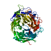
| Structure viewer | Molecule: MolmilJmol/JSmol |
|---|
- Downloads & links
-Download
| PDBx/mmCIF format | 6m0e.cif.gz | 127.4 KB | Display | PDBx/mmCIF format |
|---|---|---|---|---|
| PDB format | pdb6m0e.ent.gz | 92.3 KB | Display | PDB format |
| PDBx/mmJSON format | 6m0e.json.gz | Tree view | PDBx/mmJSON format | |
| Others |  Other downloads Other downloads |
-Validation report
| Arichive directory | https://data.pdbj.org/pub/pdb/validation_reports/m0/6m0eftp://data.pdbj.org/pub/pdb/validation_reports/m0/6m0e | HTTPS FTP |
|---|
-Related structure data
| Related structure data | 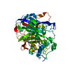 6m0dSC S: Starting model for refinement C: citing same article ( |
|---|---|
| Similar structure data |

-Links
 PDBj
PDBj

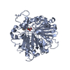
- Assembly
| Deposited unit | 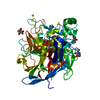
| ||||||||
|---|---|---|---|---|---|---|---|---|---|
| 1 |
| ||||||||
| Unit cell |
|
-Components
| #1: Protein | Mass: 57004.793 Da / Num. of mol.: 1 / Mutation: 198-202, 522-534 deletion Source method: isolated from a genetically manipulated source Source: (gene. exp.) Beijerinckia indica subsp. indica (strain ATCC 9039 / DSM 1715 / NCIB 8712) (bacteria)Strain: ATCC 9039 / DSM 1715 / NCIB 8712 / Gene: Bind_2021 / Production host: Escherichia coli (E. coli) / References: UniProt: B2IF78, levansucrase | ||||||||||
|---|---|---|---|---|---|---|---|---|---|---|---|
| #2: Chemical |   Mass: 24.305 Da / Num. of mol.: 3 / Source method: obtained synthetically / Formula: Mg / Feature type: SUBJECT OF INVESTIGATION Mass: 24.305 Da / Num. of mol.: 3 / Source method: obtained synthetically / Formula: Mg / Feature type: SUBJECT OF INVESTIGATION#3: Sugar | ChemComp-FRU / | Fructose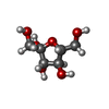  Type: D-saccharide, beta linking / Mass: 180.156 Da / Num. of mol.: 1 Type: D-saccharide, beta linking / Mass: 180.156 Da / Num. of mol.: 1Source method: isolated from a genetically manipulated source Formula: C6H12O6 / Feature type: SUBJECT OF INVESTIGATION #4: Sugar | ChemComp-BDF / Fructose  Type: D-saccharide, beta linking / Mass: 180.156 Da / Num. of mol.: 4 Type: D-saccharide, beta linking / Mass: 180.156 Da / Num. of mol.: 4Source method: isolated from a genetically manipulated source Formula: C6H12O6 / Feature type: SUBJECT OF INVESTIGATION #5: Water | ChemComp-HOH / | Water Mass: 18.015 Da / Num. of mol.: 583 / Source method: isolated from a natural source / Formula: H2O Mass: 18.015 Da / Num. of mol.: 583 / Source method: isolated from a natural source / Formula: H2OHas ligand of interest | Y | Sequence details | The gene was cloned using B. indica subsp. indica strain NBRC 3744, and DNA primers for the PCR ...The gene was cloned using B. indica subsp. indica strain NBRC 3744, and DNA primers for the PCR amplification was designed using the sequence of Bind_2021 of B. indica subsp. indica strain ATCC 9039. Two amino acid residues are different between the strain NBRC 3744 (Pro479 and Gly495) and the strain ATCC 9039 (Thr479 and Arg495). Sequence DDBJ accession number LC522529. | |
-Experimental details
-Experiment
| Experiment | Method: X-RAY DIFFRACTION / Number of used crystals: 1 |
|---|
- Sample preparation
| Crystal | Density Matthews: 1.93 Å3/Da / Density % sol: 36.14 % |
|---|---|
| Crystal grow | Temperature: 293 K / Method: vapor diffusion, hanging drop / pH: 8 Details: 30% (w/v) polyethylene glycol 10000, 0.2 M MgCl2, 0.1 M Na K tartrate, 0.1 M Tris-HCl |
-Data collection
| Diffraction | Mean temperature: 100 K / Serial crystal experiment: N |
|---|---|
| Diffraction source | Source: SYNCHROTRON / Site: Photon Factory  / Beamline: BL-17A / Wavelength: 0.98 Å / Beamline: BL-17A / Wavelength: 0.98 Å |
| Detector | Type: DECTRIS EIGER X 16M / Detector: PIXEL / Date: Dec 1, 2018 |
| Radiation | Protocol: SINGLE WAVELENGTH / Monochromatic (M) / Laue (L): M / Scattering type: x-ray |
| Radiation wavelength | Wavelength: 0.98 Å / Relative weight: 1 |
| Reflection | Resolution: 1.35→45.2 Å / Num. obs: 96105 / % possible obs: 98.9 % / Redundancy: 6.6 % / Rmerge(I) obs: 0.047 / Rpim(I) all: 0.02 / Net I/σ(I): 19.4 |
| Reflection shell | Resolution: 1.35→1.42 Å / Redundancy: 6.9 % / Rmerge(I) obs: 0.468 / Mean I/σ(I) obs: 4.1 / Num. unique obs: 13691 / Rpim(I) all: 0.19 / % possible all: 97.7 |
- Processing
| Software |
| ||||||||||||||||||||||||||||||||||||||||||||||||||||||||||||||||||||||||||||||||||||||||||||||||||||||||||||||||||||||||||||||||||||||||||||||||||||||
|---|---|---|---|---|---|---|---|---|---|---|---|---|---|---|---|---|---|---|---|---|---|---|---|---|---|---|---|---|---|---|---|---|---|---|---|---|---|---|---|---|---|---|---|---|---|---|---|---|---|---|---|---|---|---|---|---|---|---|---|---|---|---|---|---|---|---|---|---|---|---|---|---|---|---|---|---|---|---|---|---|---|---|---|---|---|---|---|---|---|---|---|---|---|---|---|---|---|---|---|---|---|---|---|---|---|---|---|---|---|---|---|---|---|---|---|---|---|---|---|---|---|---|---|---|---|---|---|---|---|---|---|---|---|---|---|---|---|---|---|---|---|---|---|---|---|---|---|---|---|---|---|
| Refinement | Method to determine structure: MOLECULAR REPLACEMENT Starting model: 6M0D Resolution: 1.35→45.2 Å / Cor.coef. Fo:Fc: 0.976 / Cor.coef. Fo:Fc free: 0.973 / Cross valid method: THROUGHOUT / ESU R: 0.05 / ESU R Free: 0.05 Details: Hydrogens have been added in their riding positions
| ||||||||||||||||||||||||||||||||||||||||||||||||||||||||||||||||||||||||||||||||||||||||||||||||||||||||||||||||||||||||||||||||||||||||||||||||||||||
| Solvent computation | Ion probe radii: 0.8 Å / Shrinkage radii: 0.8 Å / VDW probe radii: 1.2 Å | ||||||||||||||||||||||||||||||||||||||||||||||||||||||||||||||||||||||||||||||||||||||||||||||||||||||||||||||||||||||||||||||||||||||||||||||||||||||
| Displacement parameters | Biso mean: 15.306 Å2
| ||||||||||||||||||||||||||||||||||||||||||||||||||||||||||||||||||||||||||||||||||||||||||||||||||||||||||||||||||||||||||||||||||||||||||||||||||||||
| Refinement step | Cycle: LAST / Resolution: 1.35→45.2 Å
| ||||||||||||||||||||||||||||||||||||||||||||||||||||||||||||||||||||||||||||||||||||||||||||||||||||||||||||||||||||||||||||||||||||||||||||||||||||||
| Refine LS restraints |
| ||||||||||||||||||||||||||||||||||||||||||||||||||||||||||||||||||||||||||||||||||||||||||||||||||||||||||||||||||||||||||||||||||||||||||||||||||||||
| LS refinement shell |
|
