 Movie
Movie Controller

[English] 日本語
 Yorodumi
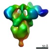
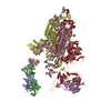
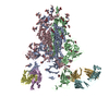
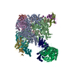
- PDB-6m04: Structure of the human homo-hexameric LRRC8D channel at 4.36 Angstroms -
+ Open data

- Basic information
| Entry | Database: PDB / ID: 6m04 | |||||||||
|---|---|---|---|---|---|---|---|---|---|---|
| Title | Structure of the human homo-hexameric LRRC8D channel at 4.36 Angstroms | |||||||||
 Components Components | Volume-regulated anion channel subunit LRRC8D | |||||||||
 Keywords Keywords | MEMBRANE PROTEIN / Ion channel | |||||||||
| Function / homology |  Function and homology information Function and homology informationMiscellaneous transport and binding events / volume-sensitive anion channel activity / taurine transmembrane transport / monoatomic anion transmembrane transport / aspartate transmembrane transport / cellular response to osmotic stress / protein hexamerization / monoatomic ion channel complex / intracellular glucose homeostasis / plasma membrane => GO:0005886 ...Miscellaneous transport and binding events / volume-sensitive anion channel activity / taurine transmembrane transport / monoatomic anion transmembrane transport / aspartate transmembrane transport / cellular response to osmotic stress / protein hexamerization / monoatomic ion channel complex / intracellular glucose homeostasis / plasma membrane => GO:0005886 / transmembrane transport / membrane => GO:0016020 / endoplasmic reticulum membrane / membrane / plasma membrane / cytoplasmSimilarity search - Function | |||||||||
| Biological species |  Homo sapiens (human) Homo sapiens (human) | |||||||||
| Method | ELECTRON MICROSCOPY / single particle reconstruction / cryo EM / Resolution: 4.36 Å | |||||||||
 Authors Authors | Nakamura, R. / Kasuya, G. / Yokoyama, T. / Shirouzu, M. / Ishitani, R. / Nureki, O. | |||||||||
| Funding support |  Japan, 2items Japan, 2items
| |||||||||
 Citation Citation | Journal: Commun Biol / Year: 2020 Title: Cryo-EM structure of the volume-regulated anion channel LRRC8D isoform identifies features important for substrate permeation. Authors: Ryoki Nakamura / Tomohiro Numata / Go Kasuya / Takeshi Yokoyama / Tomohiro Nishizawa / Tsukasa Kusakizako / Takafumi Kato / Tatsuya Hagino / Naoshi Dohmae / Masato Inoue / Kengo Watanabe / ...Authors: Ryoki Nakamura / Tomohiro Numata / Go Kasuya / Takeshi Yokoyama / Tomohiro Nishizawa / Tsukasa Kusakizako / Takafumi Kato / Tatsuya Hagino / Naoshi Dohmae / Masato Inoue / Kengo Watanabe / Hidenori Ichijo / Masahide Kikkawa / Mikako Shirouzu / Thomas J Jentsch / Ryuichiro Ishitani / Yasunobu Okada / Osamu Nureki /  Abstract: Members of the leucine-rich repeat-containing 8 (LRRC8) protein family, composed of the five LRRC8A-E isoforms, are pore-forming components of the volume-regulated anion channel (VRAC). LRRC8A and at ...Members of the leucine-rich repeat-containing 8 (LRRC8) protein family, composed of the five LRRC8A-E isoforms, are pore-forming components of the volume-regulated anion channel (VRAC). LRRC8A and at least one of the other LRRC8 isoforms assemble into heteromers to generate VRAC transport activities. Despite the availability of the LRRC8A structures, the structural basis of how LRRC8 isoforms other than LRRC8A contribute to the functional diversity of VRAC has remained elusive. Here, we present the structure of the human LRRC8D isoform, which enables the permeation of organic substrates through VRAC. The LRRC8D homo-hexamer structure displays a two-fold symmetric arrangement, and together with a structure-based electrophysiological analysis, revealed two key features. The pore constriction on the extracellular side is wider than that in the LRRC8A structures, which may explain the increased permeability of organic substrates. Furthermore, an N-terminal helix protrudes into the pore from the intracellular side and may be critical for gating. | |||||||||
| History |
|
- Structure visualization
| Movie |
Movie viewer |
|---|---|
| Structure viewer | Molecule: MolmilJmol/JSmol |
- Downloads & links
-Download
| PDBx/mmCIF format | 6m04.cif.gz | 751.4 KB | Display | PDBx/mmCIF format |
|---|---|---|---|---|
| PDB format | pdb6m04.ent.gz | 630.3 KB | Display | PDB format |
| PDBx/mmJSON format | 6m04.json.gz | Tree view | PDBx/mmJSON format | |
| Others |  Other downloads Other downloads |
-Validation report
| Arichive directory | https://data.pdbj.org/pub/pdb/validation_reports/m0/6m04ftp://data.pdbj.org/pub/pdb/validation_reports/m0/6m04 | HTTPS FTP |
|---|
-Related structure data
| Related structure data | 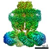 30029MC M: map data used to model this data C: citing same article ( |
|---|---|
| Similar structure data | |
| EM raw data | EMPIAR-10383 (Title: CryoEM structure of human LRRC8D / Data size: 1.7 TB Data #1: Unaligned movies of human LRRC8D isoform [micrographs - multiframe]) |

-Links
 PDBj
PDBj


- Assembly
| Deposited unit | 
|
|---|---|
| 1 |
|
-Components
| #1: Protein | / Leucine-rich repeat-containing protein 5 / Leucine-rich repeat-containing protein 8D Mass: 99311.180 Da / Num. of mol.: 6 / Mutation: L810F Source method: isolated from a genetically manipulated source Source: (gene. exp.) Homo sapiens (human) / Gene: LRRC8D, LRRC5, UNQ213/PRO239 / Cell line (production host): HEK293S / Production host: Homo sapiens (human) / Variant (production host): GntI- / References: UniProt: Q7L1W4 |
|---|
-Experimental details
-Experiment
| Experiment | Method: ELECTRON MICROSCOPY |
|---|---|
| EM experiment | Aggregation state: PARTICLE / 3D reconstruction method: single particle reconstruction |
- Sample preparation
| Component | Name: Hexameric channel of LRC8D_HUMAN / Type: COMPLEX / Entity ID: all / Source: MULTIPLE SOURCES | |||||||||||||||||||||||||
|---|---|---|---|---|---|---|---|---|---|---|---|---|---|---|---|---|---|---|---|---|---|---|---|---|---|---|
| Molecular weight | Experimental value: NO | |||||||||||||||||||||||||
| Source (natural) | Organism: Homo sapiens (human) | |||||||||||||||||||||||||
| Source (recombinant) | Organism: Homo sapiens (human) / Cell: HEK293S | |||||||||||||||||||||||||
| Buffer solution | pH: 8 Details: The solution was freshly prepared to avoid digitonin precipitation. | |||||||||||||||||||||||||
| Buffer component |
| |||||||||||||||||||||||||
| Specimen | Conc.: 3 mg/ml / Embedding applied: NO / Shadowing applied: NO / Staining applied: NO / Vitrification applied: YES / Details: This sample was monodisperse | |||||||||||||||||||||||||
| Specimen support | Grid material: COPPER/RHODIUM / Grid mesh size: 300 divisions/in. / Grid type: Quantifoil R1.2/1.3 | |||||||||||||||||||||||||
| Vitrification | Instrument: FEI VITROBOT MARK IV / Cryogen name: ETHANE / Humidity: 100 % / Chamber temperature: 277 K / Details: Blotted for 4 seconds before plunging. |
- Electron microscopy imaging
| Experimental equipment |  Model: Talos Arctica / Image courtesy: FEI Company |
|---|---|
| Microscopy | Model: FEI TALOS ARCTICA Details: Specimen holder is FEI Talos Arctica autogrid holder. |
| Electron gun | Electron source: FIELD EMISSION GUN / Accelerating voltage: 200 kV / Illumination mode: FLOOD BEAM |
| Electron lens | Mode: BRIGHT FIELDBright-field microscopy / Nominal magnification: 23500 X / Nominal defocus max: 2500 nm / Nominal defocus min: 500 nm / Cs: 2.7 mm / C2 aperture diameter: 50 µm / Alignment procedure: COMA FREE |
| Specimen holder | Cryogen: NITROGEN / Specimen holder model: OTHER / Temperature (max): 79.55 K / Temperature (min): 79.55 K |
| Image recording | Average exposure time: 15 sec. / Electron dose: 50 e/Å2 / Detector mode: SUPER-RESOLUTION / Film or detector model: GATAN K2 SUMMIT (4k x 4k) / Num. of real images: 3397 |
| Image scans | Movie frames/image: 40 / Used frames/image: 1-40 |
- Processing
| Software | Name: PHENIX / Version: 1.12_2829: / Classification: refinement | ||||||||||||||||||||||||||||||||
|---|---|---|---|---|---|---|---|---|---|---|---|---|---|---|---|---|---|---|---|---|---|---|---|---|---|---|---|---|---|---|---|---|---|
| EM software |
| ||||||||||||||||||||||||||||||||
| CTF correction | Type: PHASE FLIPPING AND AMPLITUDE CORRECTION | ||||||||||||||||||||||||||||||||
| Symmetry | Point symmetry: C2 (2 fold cyclic) | ||||||||||||||||||||||||||||||||
| 3D reconstruction | Resolution: 4.36 Å / Resolution method: FSC 0.143 CUT-OFF / Num. of particles: 247154 / Num. of class averages: 2 / Symmetry type: POINT | ||||||||||||||||||||||||||||||||
| Atomic model building | Protocol: AB INITIO MODEL / Space: REAL | ||||||||||||||||||||||||||||||||
| Refine LS restraints |
|
