 Movie
Movie Controller

[English] 日本語
 Yorodumi
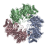
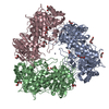
- PDB-6lvw: Polyextremophilic Beta-galactosidase from the Antarctic haloarcha... -
+ Open data

- Basic information
| Entry | Database: PDB / ID: 6lvw | ||||||
|---|---|---|---|---|---|---|---|
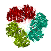
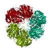
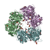
| Title | Polyextremophilic Beta-galactosidase from the Antarctic haloarchaeon Halorubrum lacusprofundi | ||||||
 Components Components | Beta-galactosidase Bga | ||||||
 Keywords Keywords |  HYDROLASE / polyextremophilic enzyme / halophile / psychrophile / extremozyme / extremophile / SUGAR BINDING PROTEIN HYDROLASE / polyextremophilic enzyme / halophile / psychrophile / extremozyme / extremophile / SUGAR BINDING PROTEIN | ||||||
| Function / homology |  Function and homology informationbeta-galactosidase complex / beta-galactosidase / beta-galactosidase activity / carbohydrate metabolic process / metal ion binding Function and homology informationbeta-galactosidase complex / beta-galactosidase / beta-galactosidase activity / carbohydrate metabolic process / metal ion bindingSimilarity search - Function | ||||||
| Biological species | Halorubrum lacusprofundi | ||||||
| Method | X-RAY DIFFRACTION / SYNCHROTRON / MOLECULAR REPLACEMENT / Resolution: 2.493 Å | ||||||
 Authors Authors | Muhammad, R. / Arold, S.T. | ||||||
 Citation Citation | Journal: Microorganisms / Year: 2020 Title: Understanding High-Salt and Cold Adaptation of a Polyextremophilic Enzyme. Authors: Karan, R. / Mathew, S. / Muhammad, R. / Bautista, D.B. / Vogler, M. / Eppinger, J. / Oliva, R. / Cavallo, L. / Arold, S.T. / Rueping, M. | ||||||
| History |
|
- Structure visualization
| Structure viewer | Molecule: MolmilJmol/JSmol |
|---|
- Downloads & links
-Download
| PDBx/mmCIF format | 6lvw.cif.gz | 281.1 KB | Display | PDBx/mmCIF format |
|---|---|---|---|---|
| PDB format | pdb6lvw.ent.gz | 228.6 KB | Display | PDB format |
| PDBx/mmJSON format | 6lvw.json.gz | Tree view | PDBx/mmJSON format | |
| Others |  Other downloads Other downloads |
-Validation report
| Arichive directory | https://data.pdbj.org/pub/pdb/validation_reports/lv/6lvwftp://data.pdbj.org/pub/pdb/validation_reports/lv/6lvw | HTTPS FTP |
|---|
-Related structure data
| Related structure data |  1kwgS S: Starting model for refinement |
|---|---|
| Similar structure data |


-Links
 PDBj
PDBj


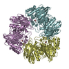
- Assembly
| Deposited unit | 
| ||||||||
|---|---|---|---|---|---|---|---|---|---|
| 1 | 
| ||||||||
| Unit cell |
|
-Components
| #1: Protein | Mass: 78136.375 Da / Num. of mol.: 1 Source method: isolated from a genetically manipulated source Source: (gene. exp.)  Halorubrum lacusprofundi (strain ATCC 49239 / DSM 5036 / JCM 8891 / ACAM 34) (archaea) Halorubrum lacusprofundi (strain ATCC 49239 / DSM 5036 / JCM 8891 / ACAM 34) (archaea)Strain: ATCC 49239 / DSM 5036 / JCM 8891 / ACAM 34 / Gene: Hlac_2868 / Production host: Haloferax volcanii (archaea) / References: UniProt: B9LW38, beta-galactosidase |
|---|---|
| #2: Chemical | ChemComp-ZN /   Mass: 65.409 Da / Num. of mol.: 1 / Source method: obtained synthetically / Formula: Zn Mass: 65.409 Da / Num. of mol.: 1 / Source method: obtained synthetically / Formula: Zn |
| #3: Chemical | ChemComp-MG /   Mass: 24.305 Da / Num. of mol.: 1 / Source method: obtained synthetically / Formula: Mg Mass: 24.305 Da / Num. of mol.: 1 / Source method: obtained synthetically / Formula: Mg |
| #4: Water | ChemComp-HOH / Water Mass: 18.015 Da / Num. of mol.: 3 / Source method: isolated from a natural source / Formula: H2O Mass: 18.015 Da / Num. of mol.: 3 / Source method: isolated from a natural source / Formula: H2O |
| Has ligand of interest | N |
-Experimental details
-Experiment
| Experiment | Method: X-RAY DIFFRACTION / Number of used crystals: 1 |
|---|
- Sample preparation
| Crystal | Density Matthews: 2.55 Å3/Da / Density % sol: 51.68 % |
|---|---|
| Crystal grow | Temperature: 298 K / Method: vapor diffusion, sitting drop / pH: 8.5 Details: 85 mM Tris-HCl (pH 8.5), 142 mM MgCl2, 2.78 M 1,6-hexanediol, 450 mM NDSB-195 |
-Data collection
| Diffraction | Mean temperature: 100 K / Serial crystal experiment: N | ||||||||||||||||||||||||||||||
|---|---|---|---|---|---|---|---|---|---|---|---|---|---|---|---|---|---|---|---|---|---|---|---|---|---|---|---|---|---|---|---|
| Diffraction source | Source: SYNCHROTRON / Site: SOLEIL  / Beamline: PROXIMA 1 / Wavelength: 0.97857 Å / Beamline: PROXIMA 1 / Wavelength: 0.97857 Å | ||||||||||||||||||||||||||||||
| Detector | Type: DECTRIS PILATUS3 S 6M / Detector: PIXEL / Date: Apr 8, 2018 | ||||||||||||||||||||||||||||||
| Radiation | Protocol: SINGLE WAVELENGTH / Monochromatic (M) / Laue (L): M / Scattering type: x-ray | ||||||||||||||||||||||||||||||
| Radiation wavelength | Wavelength: 0.97857 Å / Relative weight: 1 | ||||||||||||||||||||||||||||||
| Reflection | Resolution: 2.49→50.07 Å / Num. obs: 27127 / % possible obs: 99.3 % / Redundancy: 19.9 % / CC1/2: 0.999 / Rmerge(I) obs: 0.149 / Rpim(I) all: 0.034 / Rrim(I) all: 0.153 / Net I/σ(I): 15 / Num. measured all: 539186 / Scaling rejects: 7 | ||||||||||||||||||||||||||||||
| Reflection shell | Diffraction-ID: 1
|
- Processing
| Software |
| ||||||||||||||||||||||||||||||||||||||||||||||||||||||||||||
|---|---|---|---|---|---|---|---|---|---|---|---|---|---|---|---|---|---|---|---|---|---|---|---|---|---|---|---|---|---|---|---|---|---|---|---|---|---|---|---|---|---|---|---|---|---|---|---|---|---|---|---|---|---|---|---|---|---|---|---|---|---|
| Refinement | Method to determine structure: MOLECULAR REPLACEMENT Starting model: 1KWG Resolution: 2.493→34.357 Å / SU ML: 0.59 / Cross valid method: THROUGHOUT / σ(F): 0.01 / Phase error: 44.65
| ||||||||||||||||||||||||||||||||||||||||||||||||||||||||||||
| Solvent computation | Shrinkage radii: 0.9 Å / VDW probe radii: 1.11 Å | ||||||||||||||||||||||||||||||||||||||||||||||||||||||||||||
| Displacement parameters | Biso max: 251.44 Å2 / Biso mean: 100.9852 Å2 / Biso min: 39.8 Å2 | ||||||||||||||||||||||||||||||||||||||||||||||||||||||||||||
| Refinement step | Cycle: final / Resolution: 2.493→34.357 Å
| ||||||||||||||||||||||||||||||||||||||||||||||||||||||||||||
| LS refinement shell | Refine-ID: X-RAY DIFFRACTION / Rfactor Rfree error: 0
| ||||||||||||||||||||||||||||||||||||||||||||||||||||||||||||
| Refinement TLS params. | Method: refined / Origin x: 18.2566 Å / Origin y: 37.2514 Å / Origin z: 2.3014 Å
| ||||||||||||||||||||||||||||||||||||||||||||||||||||||||||||
| Refinement TLS group | Selection details: all |
