 Movie
Movie Controller


+ Open data

- Basic information
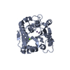
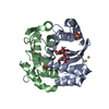
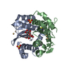
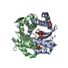
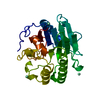
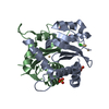
| Entry | Database: PDB / ID: 6lug | ||||||
|---|---|---|---|---|---|---|---|
| Title | Crystal structure of N(omega)-hydroxy-L-arginine hydrolase | ||||||
 Components Components | N(omega)-hydroxy-L-arginine amidinohydrolase | ||||||
 Keywords Keywords |  HYDROLASE HYDROLASE | ||||||
| Function / homology |  Function and homology information Function and homology informationNomega-hydroxy-L-arginine amidinohydrolase / hydrolase activity, acting on carbon-nitrogen (but not peptide) bonds, in linear amidines / antibiotic biosynthetic process / metal ion bindingSimilarity search - Function | ||||||
| Biological species |  Streptomyces lavendulae (bacteria) Streptomyces lavendulae (bacteria) | ||||||
| Method | X-RAY DIFFRACTION / SYNCHROTRON / SAD / MAD / Resolution: 1.9 Å | ||||||
 Authors Authors | Oda, K. / Matoba, Y. | ||||||
 Citation Citation | Journal: Acta Crystallogr D Struct Biol / Year: 2020 Title: Crystal structure of an Nomega-hydroxy-L-arginine hydrolase found in the D-cycloserine biosynthetic pathway. Authors: Oda, K. / Shimotani, N. / Kuroda, T. / Matoba, Y. | ||||||
| History |
|
- Structure visualization
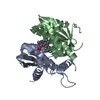
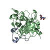
| Structure viewer | Molecule: MolmilJmol/JSmol |
|---|
- Downloads & links
-Download
| PDBx/mmCIF format | 6lug.cif.gz | 123.8 KB | Display | PDBx/mmCIF format |
|---|---|---|---|---|
| PDB format | pdb6lug.ent.gz | 94.8 KB | Display | PDB format |
| PDBx/mmJSON format | 6lug.json.gz | Tree view | PDBx/mmJSON format | |
| Others |  Other downloads Other downloads |
-Validation report
| Arichive directory | https://data.pdbj.org/pub/pdb/validation_reports/lu/6lugftp://data.pdbj.org/pub/pdb/validation_reports/lu/6lug | HTTPS FTP |
|---|
-Related structure data


-Links
 PDBj
PDBj- Assembly
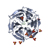
| Deposited unit | 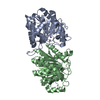
| ||||||||
|---|---|---|---|---|---|---|---|---|---|
| 1 | 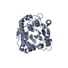
| ||||||||
| 2 | 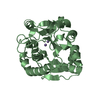
| ||||||||
| Unit cell |
|
-Components
| #1: Protein | Mass: 30016.701 Da / Num. of mol.: 2 / Fragment: N(omega)-hydroxy-L-arginine amidinohydrolase Source method: isolated from a genetically manipulated source Source: (gene. exp.) Streptomyces lavendulae (bacteria) / Gene: dcsB / Plasmid: pET21 / Production host: Escherichia coli BL21(DE3) (bacteria)References: UniProt: D2Z025, Nomega-hydroxy-L-arginine amidinohydrolase #2: Chemical | ChemComp-MN /   Mass: 54.938 Da / Num. of mol.: 4 / Source method: obtained synthetically / Formula: Mn / Feature type: SUBJECT OF INVESTIGATION Mass: 54.938 Da / Num. of mol.: 4 / Source method: obtained synthetically / Formula: Mn / Feature type: SUBJECT OF INVESTIGATION#3: Water | ChemComp-HOH / | Water Mass: 18.015 Da / Num. of mol.: 428 / Source method: isolated from a natural source / Formula: H2O Mass: 18.015 Da / Num. of mol.: 428 / Source method: isolated from a natural source / Formula: H2OHas ligand of interest | Y | |
|---|
-Experimental details
-Experiment
| Experiment | Method: X-RAY DIFFRACTION / Number of used crystals: 1 |
|---|
- Sample preparation
| Crystal | Density Matthews: 2.02 Å3/Da / Density % sol: 38.98 % |
|---|---|
| Crystal grow | Temperature: 298 K / Method: vapor diffusion, sitting drop / pH: 8.3 / Details: PEG 4000, Magnesium chloride |
-Data collection
| Diffraction | Mean temperature: 100 K / Serial crystal experiment: N |
|---|---|
| Diffraction source | Source: SYNCHROTRON / Site: SPring-8  / Beamline: BL26B1 / Wavelength: 1.8924 Å / Beamline: BL26B1 / Wavelength: 1.8924 Å |
| Detector | Type: DECTRIS EIGER X 4M / Detector: PIXEL / Date: Apr 18, 2019 |
| Radiation | Protocol: SINGLE WAVELENGTH / Monochromatic (M) / Laue (L): M / Scattering type: x-ray |
| Radiation wavelength | Wavelength: 1.8924 Å / Relative weight: 1 |
| Reflection | Resolution: 1.9→44.1 Å / Num. obs: 35027 / % possible obs: 95.3 % / Redundancy: 12.4 % / CC1/2: 0.995 / Rmerge(I) obs: 0.106 / Rpim(I) all: 0.03 / Rrim(I) all: 0.11 / Net I/σ(I): 19 / Num. measured all: 434720 / Scaling rejects: 614 |
| Reflection shell | Resolution: 1.9→1.95 Å / Redundancy: 5.8 % / Rmerge(I) obs: 0.288 / Num. unique obs: 1975 / CC1/2: 0.942 / Rpim(I) all: 0.131 / Rrim(I) all: 0.318 / % possible all: 79.2 |
-Phasing
| Phasing | Method: MAD |
|---|
- Processing
| Software |
| ||||||||||||||||||||||||||||||||||||||||||||||||||||||||||||||||||||||||||||||||||||||||||||||||||
|---|---|---|---|---|---|---|---|---|---|---|---|---|---|---|---|---|---|---|---|---|---|---|---|---|---|---|---|---|---|---|---|---|---|---|---|---|---|---|---|---|---|---|---|---|---|---|---|---|---|---|---|---|---|---|---|---|---|---|---|---|---|---|---|---|---|---|---|---|---|---|---|---|---|---|---|---|---|---|---|---|---|---|---|---|---|---|---|---|---|---|---|---|---|---|---|---|---|---|---|
| Refinement | Method to determine structure: SAD / Resolution: 1.9→44.1 Å / SU ML: 0.27 / Cross valid method: THROUGHOUT / σ(F): 1.98 / Phase error: 28.07 / Stereochemistry target values: ML
| ||||||||||||||||||||||||||||||||||||||||||||||||||||||||||||||||||||||||||||||||||||||||||||||||||
| Solvent computation | Shrinkage radii: 0.9 Å / VDW probe radii: 1.11 Å / Solvent model: FLAT BULK SOLVENT MODEL | ||||||||||||||||||||||||||||||||||||||||||||||||||||||||||||||||||||||||||||||||||||||||||||||||||
| Displacement parameters | Biso max: 78.84 Å2 / Biso mean: 19.9904 Å2 / Biso min: 6.8 Å2 | ||||||||||||||||||||||||||||||||||||||||||||||||||||||||||||||||||||||||||||||||||||||||||||||||||
| Refinement step | Cycle: final / Resolution: 1.9→44.1 Å
| ||||||||||||||||||||||||||||||||||||||||||||||||||||||||||||||||||||||||||||||||||||||||||||||||||
| LS refinement shell | Refine-ID: X-RAY DIFFRACTION / Rfactor Rfree error: 0 / Total num. of bins used: 13
|
