 Movie
Movie Controller

[English] 日本語
 Yorodumi
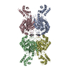
- PDB-6lox: Crystal Structure of human glutaminase with macrocyclic inhibitor -
+ Open data

- Basic information
| Entry | Database: PDB / ID: 6lox | |||||||||
|---|---|---|---|---|---|---|---|---|---|---|
| Title | Crystal Structure of human glutaminase with macrocyclic inhibitor | |||||||||
 Components Components | Glutaminase kidney isoform, mitochondrial | |||||||||
 Keywords Keywords |  PROTEIN BINDING / protein-inhibitor complex PROTEIN BINDING / protein-inhibitor complex | |||||||||
| Function / homology |  Function and homology information Function and homology informationglutamine catabolic process / regulation of respiratory gaseous exchange by nervous system process / glutamate biosynthetic process / Glutamate and glutamine metabolism / intracellular glutamate homeostasis / Glutamate Neurotransmitter Release Cycle / glutaminase / glutaminase activity / suckling behavior / TP53 Regulates Metabolic Genes ...glutamine catabolic process / regulation of respiratory gaseous exchange by nervous system process / glutamate biosynthetic process / Glutamate and glutamine metabolism / intracellular glutamate homeostasis / Glutamate Neurotransmitter Release Cycle / glutaminase / glutaminase activity / suckling behavior / TP53 Regulates Metabolic Genes / chemical synaptic transmission / protein homotetramerization / mitochondrial matrix / synapse / mitochondrion / cytosolSimilarity search - Function | |||||||||
| Biological species |  Homo sapiens (human) Homo sapiens (human) | |||||||||
| Method | X-RAY DIFFRACTION / SYNCHROTRON / MOLECULAR REPLACEMENT / Resolution: 3.2 Å | |||||||||
 Authors Authors | Bian, J. / Li, Z. / Xu, X. / Wang, J. / Li, L. | |||||||||
| Funding support |  China, 2items China, 2items
| |||||||||
 Citation Citation | Journal: J.Med.Chem. / Year: 2021 Title: Structure-Enabled Discovery of Novel Macrocyclic Inhibitors Targeting Glutaminase 1 Allosteric Binding Site. Authors: Xu, X. / Wang, J. / Wang, M. / Yuan, X. / Li, L. / Zhang, C. / Huang, H. / Jing, T. / Wang, C. / Tong, C. / Zhou, L. / Meng, Y. / Xu, P. / Kou, J. / Qiu, Z. / Li, Z. / Bian, J. | |||||||||
| History |
|
- Structure visualization
| Structure viewer | Molecule: MolmilJmol/JSmol |
|---|
- Downloads & links
-Download
| PDBx/mmCIF format | 6lox.cif.gz | 319 KB | Display | PDBx/mmCIF format |
|---|---|---|---|---|
| PDB format | pdb6lox.ent.gz | 251.1 KB | Display | PDB format |
| PDBx/mmJSON format | 6lox.json.gz | Tree view | PDBx/mmJSON format | |
| Others |  Other downloads Other downloads |
-Validation report
| Arichive directory | https://data.pdbj.org/pub/pdb/validation_reports/lo/6loxftp://data.pdbj.org/pub/pdb/validation_reports/lo/6lox | HTTPS FTP |
|---|
-Related structure data
| Related structure data |  3uo9S S: Starting model for refinement |
|---|---|
| Similar structure data |

-Links
 PDBj
PDBj
- Assembly
| Deposited unit | 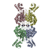
| ||||||||||||||||||||||||||||||||||||||||||||||||||||||||||||||||||||||||||||||||||||||||||||||||||||||||||||||||||||||||||||||||||||||||||||||||||||||||||||||||||||||||||||||||
|---|---|---|---|---|---|---|---|---|---|---|---|---|---|---|---|---|---|---|---|---|---|---|---|---|---|---|---|---|---|---|---|---|---|---|---|---|---|---|---|---|---|---|---|---|---|---|---|---|---|---|---|---|---|---|---|---|---|---|---|---|---|---|---|---|---|---|---|---|---|---|---|---|---|---|---|---|---|---|---|---|---|---|---|---|---|---|---|---|---|---|---|---|---|---|---|---|---|---|---|---|---|---|---|---|---|---|---|---|---|---|---|---|---|---|---|---|---|---|---|---|---|---|---|---|---|---|---|---|---|---|---|---|---|---|---|---|---|---|---|---|---|---|---|---|---|---|---|---|---|---|---|---|---|---|---|---|---|---|---|---|---|---|---|---|---|---|---|---|---|---|---|---|---|---|---|---|---|
| 1 |
| ||||||||||||||||||||||||||||||||||||||||||||||||||||||||||||||||||||||||||||||||||||||||||||||||||||||||||||||||||||||||||||||||||||||||||||||||||||||||||||||||||||||||||||||||
| Unit cell |
| ||||||||||||||||||||||||||||||||||||||||||||||||||||||||||||||||||||||||||||||||||||||||||||||||||||||||||||||||||||||||||||||||||||||||||||||||||||||||||||||||||||||||||||||||
| Noncrystallographic symmetry (NCS) | NCS domain:
NCS domain segments: Component-ID: 0 / Refine code: 0
NCS ensembles :
|
-Components
| #1: Protein | Mass: 58937.992 Da / Num. of mol.: 4 Source method: isolated from a genetically manipulated source Source: (gene. exp.) Homo sapiens (human) / Gene: GLS, GLS1, KIAA0838 / Plasmid: pET28a / Production host:  Escherichia coli BL21(DE3) (bacteria) / References: UniProt: O94925, glutaminase Escherichia coli BL21(DE3) (bacteria) / References: UniProt: O94925, glutaminase#2: Chemical |   Mass: 598.715 Da / Num. of mol.: 2 / Source method: obtained synthetically / Formula: C32H34N6O4S / Feature type: SUBJECT OF INVESTIGATION Mass: 598.715 Da / Num. of mol.: 2 / Source method: obtained synthetically / Formula: C32H34N6O4S / Feature type: SUBJECT OF INVESTIGATION#3: Water | ChemComp-HOH / | Water Mass: 18.015 Da / Num. of mol.: 13 / Source method: isolated from a natural source / Formula: H2O Mass: 18.015 Da / Num. of mol.: 13 / Source method: isolated from a natural source / Formula: H2OHas ligand of interest | Y | |
|---|
-Experimental details
-Experiment
| Experiment | Method: X-RAY DIFFRACTION / Number of used crystals: 1 |
|---|
- Sample preparation
| Crystal | Density Matthews: 2.6 Å3/Da / Density % sol: 52.77 % / Mosaicity: 0.29 ° |
|---|---|
| Crystal grow | Temperature: 289 K / Method: evaporation / pH: 8.7 / Details: PEG 6000 |
-Data collection
| Diffraction | Mean temperature: 100 K / Serial crystal experiment: N | ||||||||||||||||||||||||||||||
|---|---|---|---|---|---|---|---|---|---|---|---|---|---|---|---|---|---|---|---|---|---|---|---|---|---|---|---|---|---|---|---|
| Diffraction source | Source: SYNCHROTRON / Site: SSRF / Beamline: BL19U1 / Wavelength: 0.97853 Å | ||||||||||||||||||||||||||||||
| Detector | Type: DECTRIS PILATUS3 6M / Detector: PIXEL / Date: Oct 21, 2019 | ||||||||||||||||||||||||||||||
| Radiation | Protocol: SINGLE WAVELENGTH / Monochromatic (M) / Laue (L): M / Scattering type: x-ray | ||||||||||||||||||||||||||||||
| Radiation wavelength | Wavelength: 0.97853 Å / Relative weight: 1 | ||||||||||||||||||||||||||||||
| Reflection | Resolution: 3.2→47.14 Å / Num. obs: 39470 / % possible obs: 98.9 % / Redundancy: 3.8 % / CC1/2: 0.991 / Rmerge(I) obs: 0.15 / Rpim(I) all: 0.088 / Rrim(I) all: 0.175 / Net I/σ(I): 8.2 | ||||||||||||||||||||||||||||||
| Reflection shell | Diffraction-ID: 1
|
- Processing
| Software |
| |||||||||||||||||||||||||||||||||||||||||||||||||||||||||||||||||
|---|---|---|---|---|---|---|---|---|---|---|---|---|---|---|---|---|---|---|---|---|---|---|---|---|---|---|---|---|---|---|---|---|---|---|---|---|---|---|---|---|---|---|---|---|---|---|---|---|---|---|---|---|---|---|---|---|---|---|---|---|---|---|---|---|---|---|
| Refinement | Method to determine structure: MOLECULAR REPLACEMENT Starting model: 3UO9 Resolution: 3.2→47.14 Å / Cor.coef. Fo:Fc: 0.929 / Cor.coef. Fo:Fc free: 0.897 / Rfactor Rfree error: 0.491 / SU B: 32.658 / SU ML: 0.491 / Cross valid method: THROUGHOUT / σ(F): 0 / ESU R Free: 0.483 / Details: U VALUES : REFINED INDIVIDUALLY
| |||||||||||||||||||||||||||||||||||||||||||||||||||||||||||||||||
| Solvent computation | Ion probe radii: 0.9 Å / Shrinkage radii: 0.9 Å / VDW probe radii: 1.1 Å | |||||||||||||||||||||||||||||||||||||||||||||||||||||||||||||||||
| Displacement parameters | Biso max: 201.29 Å2 / Biso mean: 61.869 Å2 / Biso min: 18.7 Å2
| |||||||||||||||||||||||||||||||||||||||||||||||||||||||||||||||||
| Refinement step | Cycle: final / Resolution: 3.2→47.14 Å
| |||||||||||||||||||||||||||||||||||||||||||||||||||||||||||||||||
| Refine LS restraints |
| |||||||||||||||||||||||||||||||||||||||||||||||||||||||||||||||||
| Refine LS restraints NCS | Refine-ID: X-RAY DIFFRACTION / Type: interatomic distance / Weight position: 0.05
| |||||||||||||||||||||||||||||||||||||||||||||||||||||||||||||||||
| LS refinement shell | Resolution: 3.2→3.283 Å / Rfactor Rfree error: 0 / Total num. of bins used: 20
|
