 Movie
Movie Controller


+ Open data

- Basic information
| Entry | Database: PDB / ID: 6lmy | ||||||
|---|---|---|---|---|---|---|---|
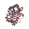
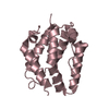
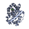
| Title | Crystal structure of DUSP22 mutant_C88S/S93A | ||||||
 Components Components | Dual specificity protein phosphatase 22 | ||||||
 Keywords Keywords |  HYDROLASE / DUSP22 / atypical DUSPs / Cysteine based protein tyrosine phosphatases (Cys-based PTPs) / active site of DUSPs HYDROLASE / DUSP22 / atypical DUSPs / Cysteine based protein tyrosine phosphatases (Cys-based PTPs) / active site of DUSPs | ||||||
| Function / homology |  Function and homology information Function and homology informationnegative regulation of non-membrane spanning protein tyrosine kinase activity / negative regulation of T cell mediated immunity / leading edge of lamellipodium / negative regulation of T cell activation / protein tyrosine/serine/threonine phosphatase activity / negative regulation of focal adhesion assembly / negative regulation of T cell receptor signaling pathway / myosin phosphatase activity / protein-serine/threonine phosphatase / filamentous actin ...negative regulation of non-membrane spanning protein tyrosine kinase activity / negative regulation of T cell mediated immunity / leading edge of lamellipodium / negative regulation of T cell activation / protein tyrosine/serine/threonine phosphatase activity / negative regulation of focal adhesion assembly / negative regulation of T cell receptor signaling pathway / myosin phosphatase activity / protein-serine/threonine phosphatase / filamentous actin / non-membrane spanning protein tyrosine phosphatase activity / peptidyl-tyrosine dephosphorylation / dephosphorylation / cellular response to epidermal growth factor stimulus / negative regulation of cell migration / transforming growth factor beta receptor signaling pathway / protein tyrosine kinase binding / protein-tyrosine-phosphatase / protein tyrosine phosphatase activity / positive regulation of JNK cascade / regulation of cell population proliferation / negative regulation of transcription by RNA polymerase II / signal transduction / plasma membrane / cytosol / cytoplasmSimilarity search - Function | ||||||
| Biological species |  Homo sapiens (human) Homo sapiens (human) | ||||||
| Method | X-RAY DIFFRACTION / SYNCHROTRON / MOLECULAR REPLACEMENT / molecular replacement / Resolution: 1.5 Å | ||||||
 Authors Authors | Lai, C.H. / Lyu, P.C. | ||||||
 Citation Citation | Journal: Int J Mol Sci / Year: 2020 Title: Structural Insights into the Active Site Formation of DUSP22 in N-loop-containing Protein Tyrosine Phosphatases. Authors: Lai, C.H. / Chang, C.C. / Chuang, H.C. / Tan, T.H. / Lyu, P.C. | ||||||
| History |
|
- Structure visualization
| Structure viewer | Molecule: MolmilJmol/JSmol |
|---|
- Downloads & links
-Download
| PDBx/mmCIF format | 6lmy.cif.gz | 48.9 KB | Display | PDBx/mmCIF format |
|---|---|---|---|---|
| PDB format | pdb6lmy.ent.gz | 31.9 KB | Display | PDB format |
| PDBx/mmJSON format | 6lmy.json.gz | Tree view | PDBx/mmJSON format | |
| Others |  Other downloads Other downloads |
-Validation report
| Arichive directory | https://data.pdbj.org/pub/pdb/validation_reports/lm/6lmyftp://data.pdbj.org/pub/pdb/validation_reports/lm/6lmy | HTTPS FTP |
|---|
-Related structure data
| Related structure data | 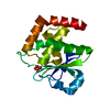 6l1sC 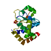 6lotC 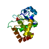 6louC 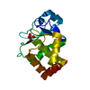 6lvqC 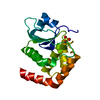 7c8sC  1wrmS  6kmi C: citing same article ( S: Starting model for refinement |
|---|---|
| Similar structure data |


-Links
 PDBj
PDBj


- Assembly
| Deposited unit | 
| ||||||||
|---|---|---|---|---|---|---|---|---|---|
| 1 |
| ||||||||
| Unit cell |
|
-Components
| #1: Protein | Mass: 17779.314 Da / Num. of mol.: 1 / Mutation: C88S,S93A Source method: isolated from a genetically manipulated source Source: (gene. exp.) Homo sapiens (human) / Gene: DUSP22, JSP1, LMWDSP2, MKPX / Production host:  Escherichia coli (E. coli) Escherichia coli (E. coli)References: UniProt: Q9NRW4, protein-serine/threonine phosphatase, protein-tyrosine-phosphatase |
|---|---|
| #2: Chemical | ChemComp-PO4 / Phosphate  Mass: 94.971 Da / Num. of mol.: 1 / Source method: obtained synthetically / Formula: PO4 / Feature type: SUBJECT OF INVESTIGATION Mass: 94.971 Da / Num. of mol.: 1 / Source method: obtained synthetically / Formula: PO4 / Feature type: SUBJECT OF INVESTIGATION |
| #3: Water | ChemComp-HOH / Water Mass: 18.015 Da / Num. of mol.: 111 / Source method: isolated from a natural source / Formula: H2O Mass: 18.015 Da / Num. of mol.: 111 / Source method: isolated from a natural source / Formula: H2O |
| Has ligand of interest | Y |
-Experimental details
-Experiment
| Experiment | Method: X-RAY DIFFRACTION / Number of used crystals: 1 |
|---|
- Sample preparation
| Crystal | Density Matthews: 2.5 Å3/Da / Density % sol: 50.82 % |
|---|---|
| Crystal grow | Temperature: 293 K / Method: vapor diffusion, hanging drop / pH: 8 Details: 0.2 M imidazole, 0.4 M NaH2PO4, 1.6 M K2HPO4, 0.2 M NaCl |
-Data collection
| Diffraction | Mean temperature: 100 K / Serial crystal experiment: N | |||||||||||||||||||||||||||||||||||||||||||||||||||||||||||||||||||||||||||||||||||||||||||||||||||
|---|---|---|---|---|---|---|---|---|---|---|---|---|---|---|---|---|---|---|---|---|---|---|---|---|---|---|---|---|---|---|---|---|---|---|---|---|---|---|---|---|---|---|---|---|---|---|---|---|---|---|---|---|---|---|---|---|---|---|---|---|---|---|---|---|---|---|---|---|---|---|---|---|---|---|---|---|---|---|---|---|---|---|---|---|---|---|---|---|---|---|---|---|---|---|---|---|---|---|---|---|
| Diffraction source | Source: SYNCHROTRON / Site: NSRRC  / Beamline: BL15A1 / Wavelength: 1 Å / Beamline: BL15A1 / Wavelength: 1 Å | |||||||||||||||||||||||||||||||||||||||||||||||||||||||||||||||||||||||||||||||||||||||||||||||||||
| Detector | Type: RAYONIX MX300HE / Detector: CCD / Date: Dec 10, 2018 | |||||||||||||||||||||||||||||||||||||||||||||||||||||||||||||||||||||||||||||||||||||||||||||||||||
| Radiation | Protocol: SINGLE WAVELENGTH / Monochromatic (M) / Laue (L): M / Scattering type: x-ray | |||||||||||||||||||||||||||||||||||||||||||||||||||||||||||||||||||||||||||||||||||||||||||||||||||
| Radiation wavelength | Wavelength: 1 Å / Relative weight: 1 | |||||||||||||||||||||||||||||||||||||||||||||||||||||||||||||||||||||||||||||||||||||||||||||||||||
| Reflection | Resolution: 1.5→30 Å / Num. obs: 28189 / % possible obs: 99.5 % / Redundancy: 3.6 % / Rmerge(I) obs: 0.056 / Rpim(I) all: 0.034 / Rrim(I) all: 0.066 / Χ2: 1.015 / Net I/σ(I): 14 | |||||||||||||||||||||||||||||||||||||||||||||||||||||||||||||||||||||||||||||||||||||||||||||||||||
| Reflection shell | Diffraction-ID: 1
|
-Phasing
| Phasing | Method: molecular replacement | |||||||||
|---|---|---|---|---|---|---|---|---|---|---|
| Phasing MR |
|
- Processing
| Software |
| ||||||||||||||||||||||||||||||||||||||||||||||||||||||||||||||||||||||||||||||||||||||||||
|---|---|---|---|---|---|---|---|---|---|---|---|---|---|---|---|---|---|---|---|---|---|---|---|---|---|---|---|---|---|---|---|---|---|---|---|---|---|---|---|---|---|---|---|---|---|---|---|---|---|---|---|---|---|---|---|---|---|---|---|---|---|---|---|---|---|---|---|---|---|---|---|---|---|---|---|---|---|---|---|---|---|---|---|---|---|---|---|---|---|---|---|
| Refinement | Method to determine structure: MOLECULAR REPLACEMENT Starting model: 1WRM Resolution: 1.5→25.61 Å / SU ML: 0.15 / Cross valid method: THROUGHOUT / σ(F): 1.36 / Phase error: 20.84
| ||||||||||||||||||||||||||||||||||||||||||||||||||||||||||||||||||||||||||||||||||||||||||
| Solvent computation | Shrinkage radii: 0.9 Å / VDW probe radii: 1.11 Å | ||||||||||||||||||||||||||||||||||||||||||||||||||||||||||||||||||||||||||||||||||||||||||
| Displacement parameters | Biso max: 51.21 Å2 / Biso mean: 22.5903 Å2 / Biso min: 9.73 Å2 | ||||||||||||||||||||||||||||||||||||||||||||||||||||||||||||||||||||||||||||||||||||||||||
| Refinement step | Cycle: final / Resolution: 1.5→25.61 Å
| ||||||||||||||||||||||||||||||||||||||||||||||||||||||||||||||||||||||||||||||||||||||||||
| LS refinement shell | Refine-ID: X-RAY DIFFRACTION / Rfactor Rfree error: 0
|
