 Movie
Movie Controller


+ Open data

- Basic information
| Entry | Database: PDB / ID: 1wrm | ||||||
|---|---|---|---|---|---|---|---|
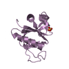
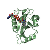
| Title | Crystal structure of JSP-1 | ||||||
 Components Components | dual specificity phosphatase 22 | ||||||
 Keywords Keywords |  HYDROLASE / phosphatase / DSP / JNK HYDROLASE / phosphatase / DSP / JNK | ||||||
| Function / homology |  Function and homology information Function and homology informationnegative regulation of non-membrane spanning protein tyrosine kinase activity / negative regulation of T cell mediated immunity / leading edge of lamellipodium / negative regulation of T cell activation / protein tyrosine/serine/threonine phosphatase activity / negative regulation of focal adhesion assembly / negative regulation of T cell receptor signaling pathway / myosin phosphatase activity / protein-serine/threonine phosphatase / filamentous actin ...negative regulation of non-membrane spanning protein tyrosine kinase activity / negative regulation of T cell mediated immunity / leading edge of lamellipodium / negative regulation of T cell activation / protein tyrosine/serine/threonine phosphatase activity / negative regulation of focal adhesion assembly / negative regulation of T cell receptor signaling pathway / myosin phosphatase activity / protein-serine/threonine phosphatase / filamentous actin / non-membrane spanning protein tyrosine phosphatase activity / peptidyl-tyrosine dephosphorylation / dephosphorylation / cellular response to epidermal growth factor stimulus / negative regulation of cell migration / transforming growth factor beta receptor signaling pathway / protein tyrosine kinase binding / protein-tyrosine-phosphatase / protein tyrosine phosphatase activity / positive regulation of JNK cascade / regulation of cell population proliferation / negative regulation of transcription by RNA polymerase II / signal transduction / plasma membrane / cytosol / cytoplasmSimilarity search - Function | ||||||
| Biological species |  Homo sapiens (human) Homo sapiens (human) | ||||||
| Method | X-RAY DIFFRACTION / SYNCHROTRON / MOLECULAR REPLACEMENT / Resolution: 1.5 Å | ||||||
 Authors Authors | Yokota, T. / Kashima, A. / Kato, R. / Sugio, S. | ||||||
 Citation Citation | Journal: Proteins / Year: 2006 Title: Crystal structure of human dual specificity phosphatase, JNK stimulatory phosphatase-1, at 1.5 A resolution Authors: Yokota, T. / Nara, Y. / Kashima, A. / Matsubara, K. / Misawa, S. / Kato, R. / Sugio, S. | ||||||
| History |
|
- Structure visualization
| Structure viewer | Molecule: MolmilJmol/JSmol |
|---|
- Downloads & links
-Download
| PDBx/mmCIF format | 1wrm.cif.gz | 45.5 KB | Display | PDBx/mmCIF format |
|---|---|---|---|---|
| PDB format | pdb1wrm.ent.gz | 31.6 KB | Display | PDB format |
| PDBx/mmJSON format | 1wrm.json.gz | Tree view | PDBx/mmJSON format | |
| Others |  Other downloads Other downloads |
-Validation report
| Arichive directory | https://data.pdbj.org/pub/pdb/validation_reports/wr/1wrmftp://data.pdbj.org/pub/pdb/validation_reports/wr/1wrm | HTTPS FTP |
|---|
-Related structure data
| Similar structure data |
|---|


-Links
 PDBj
PDBj


- Assembly
| Deposited unit | 
| ||||||||
|---|---|---|---|---|---|---|---|---|---|
| 1 |
| ||||||||
| Unit cell |
|
-Components
| #1: Protein | Mass: 18681.258 Da / Num. of mol.: 1 / Fragment: residues 1-163 Source method: isolated from a genetically manipulated source Source: (gene. exp.) Homo sapiens (human) / Plasmid: pET32-a / Production host:  Escherichia coli (E. coli) / Strain (production host): BL21 (DE4)-RP / References: UniProt: Q9NRW4, protein-tyrosine-phosphatase Escherichia coli (E. coli) / Strain (production host): BL21 (DE4)-RP / References: UniProt: Q9NRW4, protein-tyrosine-phosphatase |
|---|---|
| #2: Chemical | ChemComp-MES / MES (buffer)  Mass: 195.237 Da / Num. of mol.: 1 / Source method: obtained synthetically / Formula: C6H13NO4S / Comment: pH buffer*YM Mass: 195.237 Da / Num. of mol.: 1 / Source method: obtained synthetically / Formula: C6H13NO4S / Comment: pH buffer*YM |
| #3: Water | ChemComp-HOH / Water Mass: 18.015 Da / Num. of mol.: 80 / Source method: isolated from a natural source / Formula: H2O Mass: 18.015 Da / Num. of mol.: 80 / Source method: isolated from a natural source / Formula: H2O |
-Experimental details
-Experiment
| Experiment | Method: X-RAY DIFFRACTION / Number of used crystals: 1 |
|---|
- Sample preparation
| Crystal | Density Matthews: 2.4 Å3/Da / Density % sol: 49.4 % |
|---|---|
| Crystal grow | Temperature: 278 K / Method: vapor diffusion / pH: 6.8 Details: PEG4000, magnesium chloride, MES-NaOH, pH 6.8, VAPOR DIFFUSION, temperature 278K |
-Data collection
| Diffraction | Mean temperature: 100 K |
|---|---|
| Diffraction source | Source: SYNCHROTRON / Site: SPring-8  / Beamline: BL24XU / Wavelength: 0.83565 Å / Beamline: BL24XU / Wavelength: 0.83565 Å |
| Detector | Type: RIGAKU RAXIS V / Detector: IMAGE PLATE / Date: Oct 21, 2003 |
| Radiation | Protocol: SINGLE WAVELENGTH / Monochromatic (M) / Laue (L): M / Scattering type: x-ray |
| Radiation wavelength | Wavelength: 0.83565 Å / Relative weight: 1 |
| Reflection | Resolution: 1.5→50 Å / Num. all: 24804 / Num. obs: 24754 / % possible obs: 91.2 % / Observed criterion σ(F): 1 / Observed criterion σ(I): 1 / Redundancy: 3.3 % / Biso Wilson estimate: 18.6 Å2 / Rmerge(I) obs: 0.052 / Net I/σ(I): 37.8 |
| Reflection shell | Resolution: 1.5→1.55 Å / Redundancy: 2.7 % / Rmerge(I) obs: 0.261 / Mean I/σ(I) obs: 4.1 / % possible all: 83.9 |
- Processing
| Software |
| |||||||||||||||||||||||||
|---|---|---|---|---|---|---|---|---|---|---|---|---|---|---|---|---|---|---|---|---|---|---|---|---|---|---|
| Refinement | Method to determine structure: MOLECULAR REPLACEMENT / Resolution: 1.5→40.89 Å / Rfactor Rfree error: 0.008 / Data cutoff high absF: 1192695.44 / Data cutoff low absF: 0 / Isotropic thermal model: GROUP / Cross valid method: THROUGHOUT / σ(F): 0 / Stereochemistry target values: Engh & Huber
| |||||||||||||||||||||||||
| Solvent computation | Solvent model: FLAT MODEL / Bsol: 43.4755 Å2 / ksol: 0.361237 e/Å3 | |||||||||||||||||||||||||
| Displacement parameters | Biso mean: 30.4 Å2
| |||||||||||||||||||||||||
| Refine analyze |
| |||||||||||||||||||||||||
| Refinement step | Cycle: LAST / Resolution: 1.5→40.89 Å
| |||||||||||||||||||||||||
| Refine LS restraints |
| |||||||||||||||||||||||||
| LS refinement shell | Highest resolution: 1.5 Å / Total num. of bins used: 6 /
| |||||||||||||||||||||||||
| Xplor file |
|
