 Movie
Movie Controller


+ Open data

- Basic information
| Entry | Database: PDB / ID: 6ljf | |||||||||
|---|---|---|---|---|---|---|---|---|---|---|
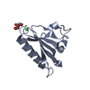
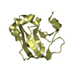
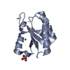
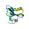
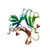
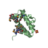
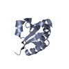
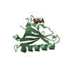
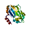
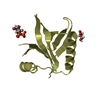
| Title | Crystal structure of gelsolin G3 domain (calcium condition) | |||||||||
 Components Components | Gelsolin | |||||||||
 Keywords Keywords | CYTOSOLIC PROTEIN / fragmin / gelsolin family protein / calcium regulation / actin filament severing | |||||||||
| Function / homology |  Function and homology information Function and homology informationstriated muscle atrophy / regulation of establishment of T cell polarity / regulation of plasma membrane raft polarization / regulation of receptor clustering / renal protein absorption / positive regulation of keratinocyte apoptotic process / positive regulation of protein processing in phagocytic vesicle / positive regulation of actin nucleation / phosphatidylinositol 3-kinase catalytic subunit binding / positive regulation of cysteine-type endopeptidase activity involved in apoptotic signaling pathway ...striated muscle atrophy / regulation of establishment of T cell polarity / regulation of plasma membrane raft polarization / regulation of receptor clustering / renal protein absorption / positive regulation of keratinocyte apoptotic process / positive regulation of protein processing in phagocytic vesicle / positive regulation of actin nucleation / phosphatidylinositol 3-kinase catalytic subunit binding / positive regulation of cysteine-type endopeptidase activity involved in apoptotic signaling pathway / actin cap / sequestering of actin monomers / regulation of podosome assembly / myosin II binding / negative regulation of viral entry into host cell / actin filament severing / actin filament capping / barbed-end actin filament capping / actin polymerization or depolymerization / actin filament depolymerization / cell projection assembly / cardiac muscle cell contraction / podosome / Sensory processing of sound by outer hair cells of the cochlea / relaxation of cardiac muscle / phagocytosis, engulfment / cortical actin cytoskeleton / hepatocyte apoptotic process / cilium assembly / sarcoplasm / Caspase-mediated cleavage of cytoskeletal proteins / phagocytic vesicle / phosphatidylinositol-4,5-bisphosphate binding / actin filament polymerization / response to muscle stretch / central nervous system development / actin filament organization / protein destabilization / cellular response to type II interferon / actin filament binding / actin cytoskeleton / lamellipodium / actin binding / secretory granule lumen / blood microparticle / ficolin-1-rich granule lumen / amyloid fibril formation / Amyloid fiber formation / focal adhesion / calcium ion binding / Neutrophil degranulation / positive regulation of gene expression / extracellular space / extracellular exosome / extracellular region / plasma membrane / cytosol / cytoplasmSimilarity search - Function | |||||||||
| Biological species |  Homo sapiens (human) Homo sapiens (human) | |||||||||
| Method | X-RAY DIFFRACTION / SYNCHROTRON / MOLECULAR REPLACEMENT / molecular replacement / Resolution: 1.5 Å | |||||||||
 Authors Authors | Takeda, S. | |||||||||
| Funding support |  Japan, 2items Japan, 2items
| |||||||||
 Citation Citation | Journal: J.Muscle Res.Cell.Motil. / Year: 2020 Title: Novel inter-domain Ca2+-binding site in the gelsolin superfamily protein fragmin. Authors: Takeda, S. / Fujiwara, I. / Sugimoto, Y. / Oda, T. / Narita, A. / Maeda, Y. | |||||||||
| History |
|
- Structure visualization
| Structure viewer | Molecule: MolmilJmol/JSmol |
|---|
- Downloads & links
-Download
| PDBx/mmCIF format | 6ljf.cif.gz | 67.7 KB | Display | PDBx/mmCIF format |
|---|---|---|---|---|
| PDB format | pdb6ljf.ent.gz | 43.3 KB | Display | PDB format |
| PDBx/mmJSON format | 6ljf.json.gz | Tree view | PDBx/mmJSON format | |
| Others |  Other downloads Other downloads |
-Validation report
| Arichive directory | https://data.pdbj.org/pub/pdb/validation_reports/lj/6ljfftp://data.pdbj.org/pub/pdb/validation_reports/lj/6ljf | HTTPS FTP |
|---|
-Related structure data
| Related structure data | 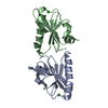 6kwzC 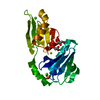 6ljcC  6ljdC  6ljeC  3ffkS S: Starting model for refinement C: citing same article ( |
|---|---|
| Similar structure data |
-Links
 PDBj
PDBj

- Assembly
| Deposited unit | 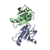
| ||||||||||||
|---|---|---|---|---|---|---|---|---|---|---|---|---|---|
| 1 | 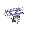
| ||||||||||||
| 2 | 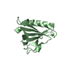
| ||||||||||||
| Unit cell |
|
-Components
| #1: Protein | / AGEL / Actin-depolymerizing factor / ADF / Brevin Mass: 11259.854 Da / Num. of mol.: 2 / Fragment: UNP residues 297-397 Source method: isolated from a genetically manipulated source Source: (gene. exp.) Homo sapiens (human) / Gene: GSN / Production host:  Escherichia coli (E. coli) / References: UniProt: P06396 Escherichia coli (E. coli) / References: UniProt: P06396#2: Chemical |   Mass: 40.078 Da / Num. of mol.: 2 / Source method: obtained synthetically / Formula: Ca Mass: 40.078 Da / Num. of mol.: 2 / Source method: obtained synthetically / Formula: Ca#3: Chemical | Glycerol  Mass: 92.094 Da / Num. of mol.: 2 / Source method: obtained synthetically / Formula: C3H8O3 Mass: 92.094 Da / Num. of mol.: 2 / Source method: obtained synthetically / Formula: C3H8O3#4: Water | ChemComp-HOH / | Water Mass: 18.015 Da / Num. of mol.: 300 / Source method: isolated from a natural source / Formula: H2O Mass: 18.015 Da / Num. of mol.: 300 / Source method: isolated from a natural source / Formula: H2OHas ligand of interest | N | |
|---|
-Experimental details
-Experiment
| Experiment | Method: X-RAY DIFFRACTION / Number of used crystals: 1 |
|---|
- Sample preparation
| Crystal | Density Matthews: 2.21 Å3/Da / Density % sol: 44.39 % |
|---|---|
| Crystal grow | Temperature: 293 K / Method: vapor diffusion, sitting drop / Details: PEG3350, ammonium sulphate, calcium chloride |
-Data collection
| Diffraction | Mean temperature: 95 K / Serial crystal experiment: N | ||||||||||||||||||||||||||||||||||||||||||||||||||||||||||||||||||||||||||||||||
|---|---|---|---|---|---|---|---|---|---|---|---|---|---|---|---|---|---|---|---|---|---|---|---|---|---|---|---|---|---|---|---|---|---|---|---|---|---|---|---|---|---|---|---|---|---|---|---|---|---|---|---|---|---|---|---|---|---|---|---|---|---|---|---|---|---|---|---|---|---|---|---|---|---|---|---|---|---|---|---|---|---|
| Diffraction source | Source: SYNCHROTRON / Site: AichiSR / Beamline: BL2S1 / Wavelength: 1.12 Å | ||||||||||||||||||||||||||||||||||||||||||||||||||||||||||||||||||||||||||||||||
| Detector | Type: ADSC QUANTUM 315r / Detector: CCD / Date: Dec 1, 2017 | ||||||||||||||||||||||||||||||||||||||||||||||||||||||||||||||||||||||||||||||||
| Radiation | Protocol: SINGLE WAVELENGTH / Monochromatic (M) / Laue (L): M / Scattering type: x-ray | ||||||||||||||||||||||||||||||||||||||||||||||||||||||||||||||||||||||||||||||||
| Radiation wavelength | Wavelength: 1.12 Å / Relative weight: 1 | ||||||||||||||||||||||||||||||||||||||||||||||||||||||||||||||||||||||||||||||||
| Reflection | Resolution: 1.5→40.809 Å / Num. obs: 31464 / % possible obs: 99.5 % / Redundancy: 4.858 % / Biso Wilson estimate: 22.829 Å2 / CC1/2: 0.999 / Rmerge(I) obs: 0.061 / Rrim(I) all: 0.069 / Χ2: 1.03 / Net I/σ(I): 16.66 | ||||||||||||||||||||||||||||||||||||||||||||||||||||||||||||||||||||||||||||||||
| Reflection shell | Diffraction-ID: 1
|
-Phasing
| Phasing | Method: molecular replacement |
|---|
- Processing
| Software |
| ||||||||||||||||||||||||||||||||||||||||||||||||||||||||||||||||||||||||||||||||||||
|---|---|---|---|---|---|---|---|---|---|---|---|---|---|---|---|---|---|---|---|---|---|---|---|---|---|---|---|---|---|---|---|---|---|---|---|---|---|---|---|---|---|---|---|---|---|---|---|---|---|---|---|---|---|---|---|---|---|---|---|---|---|---|---|---|---|---|---|---|---|---|---|---|---|---|---|---|---|---|---|---|---|---|---|---|---|
| Refinement | Method to determine structure: MOLECULAR REPLACEMENT Starting model: 3ffk Resolution: 1.5→40.809 Å / SU ML: 0.161 / Cross valid method: FREE R-VALUE / σ(F): 1.36 / Phase error: 18.7057
| ||||||||||||||||||||||||||||||||||||||||||||||||||||||||||||||||||||||||||||||||||||
| Solvent computation | Shrinkage radii: 0.9 Å / VDW probe radii: 1.11 Å | ||||||||||||||||||||||||||||||||||||||||||||||||||||||||||||||||||||||||||||||||||||
| Displacement parameters | Biso mean: 20.52 Å2 | ||||||||||||||||||||||||||||||||||||||||||||||||||||||||||||||||||||||||||||||||||||
| Refinement step | Cycle: LAST / Resolution: 1.5→40.809 Å
| ||||||||||||||||||||||||||||||||||||||||||||||||||||||||||||||||||||||||||||||||||||
| Refine LS restraints |
| ||||||||||||||||||||||||||||||||||||||||||||||||||||||||||||||||||||||||||||||||||||
| LS refinement shell |
|
