 Movie
Movie Controller


+ Open data

- Basic information
| Entry | Database: PDB / ID: 6lgj | ||||||
|---|---|---|---|---|---|---|---|
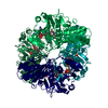
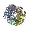
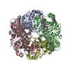
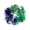
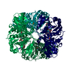
| Title | Crystal structure of an oxido-reductase | ||||||
 Components Components | Glyceraldehyde-3-phosphate dehydrogenase Glyceraldehyde 3-phosphate dehydrogenase Glyceraldehyde 3-phosphate dehydrogenase | ||||||
 Keywords Keywords | OXIDOREDUCTASE / Inhibitor | ||||||
| Function / homology |  Function and homology information Function and homology informationlate endosome lumen / Glycolysis / Gluconeogenesis / peptidyl-cysteine S-trans-nitrosylation / Transferases; Transferring nitrogenous groups; Transferring other nitrogenous groups / peptidyl-cysteine S-nitrosylase activity / negative regulation of vascular associated smooth muscle cell apoptotic process / glyceraldehyde-3-phosphate dehydrogenase (phosphorylating) / glyceraldehyde-3-phosphate dehydrogenase (NAD+) (phosphorylating) activity / glyceraldehyde-3-phosphate biosynthetic process ...late endosome lumen / Glycolysis / Gluconeogenesis / peptidyl-cysteine S-trans-nitrosylation / Transferases; Transferring nitrogenous groups; Transferring other nitrogenous groups / peptidyl-cysteine S-nitrosylase activity / negative regulation of vascular associated smooth muscle cell apoptotic process / glyceraldehyde-3-phosphate dehydrogenase (phosphorylating) / glyceraldehyde-3-phosphate dehydrogenase (NAD+) (phosphorylating) activity / glyceraldehyde-3-phosphate biosynthetic process / GAIT complex / canonical glycolysis / positive regulation of type I interferon production / postsynaptic density, intracellular component / nitric oxide mediated signal transduction / cAMP-mediated signaling / gluconeogenesis / glycolytic process / microtubule cytoskeleton organization / NAD binding / microtubule cytoskeleton / myelin sheath / NADP binding / microtubule binding / neuron apoptotic process / positive regulation of canonical NF-kappaB signal transduction / negative regulation of translation / protein stabilization / innate immune response / glutamatergic synapse / enzyme binding / mitochondrion / identical protein binding / nucleus / plasma membrane / cytosol / cytoplasmSimilarity search - Function | ||||||
| Biological species |  Mus musculus (house mouse) Mus musculus (house mouse) | ||||||
| Method | X-RAY DIFFRACTION / SYNCHROTRON / MOLECULAR REPLACEMENT / Resolution: 2.4 Å | ||||||
 Authors Authors | Yang, Y. / Lei, J. / Yin, L. | ||||||
| Funding support |  China, 1items China, 1items
| ||||||
 Citation Citation | Journal: To be published Title: Crystal structure of an oxido-reductase Authors: Yang, Y. / Lei, J. / Yin, L. | ||||||
| History |
|
- Structure visualization
| Structure viewer | Molecule: MolmilJmol/JSmol |
|---|
- Downloads & links
-Download
| PDBx/mmCIF format | 6lgj.cif.gz | 514.4 KB | Display | PDBx/mmCIF format |
|---|---|---|---|---|
| PDB format | pdb6lgj.ent.gz | 423.2 KB | Display | PDB format |
| PDBx/mmJSON format | 6lgj.json.gz | Tree view | PDBx/mmJSON format | |
| Others |  Other downloads Other downloads |
-Validation report
| Arichive directory | https://data.pdbj.org/pub/pdb/validation_reports/lg/6lgjftp://data.pdbj.org/pub/pdb/validation_reports/lg/6lgj | HTTPS FTP |
|---|
-Related structure data
| Related structure data |  4wncS S: Starting model for refinement |
|---|---|
| Similar structure data |


-Links
 PDBj
PDBj
- Assembly
| Deposited unit | 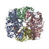
| ||||||||||
|---|---|---|---|---|---|---|---|---|---|---|---|
| 1 |
| ||||||||||
| Unit cell |
|
-Components
| #1: Protein | Glyceraldehyde 3-phosphate dehydrogenase / GAPDH / Peptidyl-cysteine S-nitrosylase GAPDH / oxido-reductase Mass: 35851.957 Da / Num. of mol.: 4 Source method: isolated from a genetically manipulated source Source: (gene. exp.) Mus musculus (house mouse) / Gene: Gapdh, Gapd / Production host:  Escherichia coli (E. coli) Escherichia coli (E. coli)References: UniProt: P16858, glyceraldehyde-3-phosphate dehydrogenase (phosphorylating), Transferases; Transferring nitrogenous groups; Transferring other nitrogenous groups#2: Chemical | ChemComp-NAD / Nicotinamide adenine dinucleotide  Mass: 663.425 Da / Num. of mol.: 4 / Source method: isolated from a natural source / Formula: C21H27N7O14P2 / Feature type: SUBJECT OF INVESTIGATION / Comment: NAD*YM Mass: 663.425 Da / Num. of mol.: 4 / Source method: isolated from a natural source / Formula: C21H27N7O14P2 / Feature type: SUBJECT OF INVESTIGATION / Comment: NAD*YM#3: Water | ChemComp-HOH / | Water Mass: 18.015 Da / Num. of mol.: 450 / Source method: isolated from a natural source / Formula: H2O Mass: 18.015 Da / Num. of mol.: 450 / Source method: isolated from a natural source / Formula: H2OHas ligand of interest | Y | |
|---|
-Experimental details
-Experiment
| Experiment | Method: X-RAY DIFFRACTION / Number of used crystals: 1 |
|---|
- Sample preparation
| Crystal | Density Matthews: 2.51 Å3/Da / Density % sol: 51.03 % |
|---|---|
| Crystal grow | Temperature: 277 K / Method: vapor diffusion, sitting drop / Details: 0.1M Tris, pH8.5, 25% PEG 3350 |
-Data collection
| Diffraction | Mean temperature: 95 K / Serial crystal experiment: N |
|---|---|
| Diffraction source | Source: SYNCHROTRON / Site: SSRF / Beamline: BL19U1 / Wavelength: 0.979 Å |
| Detector | Type: ADSC QUANTUM 315 / Detector: CCD / Date: Dec 11, 2018 |
| Radiation | Protocol: SINGLE WAVELENGTH / Monochromatic (M) / Laue (L): M / Scattering type: x-ray |
| Radiation wavelength | Wavelength: 0.979 Å / Relative weight: 1 |
| Reflection | Resolution: 2.4→47.98 Å / Num. obs: 49776 / % possible obs: 95.66 % / Redundancy: 3.1 % / Biso Wilson estimate: 29.41 Å2 / CC1/2: 0.978 / Net I/σ(I): 5.18 |
| Reflection shell | Resolution: 2.4→2.486 Å / Num. unique obs: 4985 / CC1/2: 0.927 |
- Processing
| Software |
| ||||||||||||||||||||||||||||||||||||||||||||||||||||||||||||||||||||||||||||||||||||||||||
|---|---|---|---|---|---|---|---|---|---|---|---|---|---|---|---|---|---|---|---|---|---|---|---|---|---|---|---|---|---|---|---|---|---|---|---|---|---|---|---|---|---|---|---|---|---|---|---|---|---|---|---|---|---|---|---|---|---|---|---|---|---|---|---|---|---|---|---|---|---|---|---|---|---|---|---|---|---|---|---|---|---|---|---|---|---|---|---|---|---|---|---|
| Refinement | Method to determine structure: MOLECULAR REPLACEMENT Starting model: 4WNC Resolution: 2.4→47.979 Å / SU ML: 0.25 / Cross valid method: THROUGHOUT / σ(F): 1.35 / Phase error: 22.74
| ||||||||||||||||||||||||||||||||||||||||||||||||||||||||||||||||||||||||||||||||||||||||||
| Solvent computation | Shrinkage radii: 0.9 Å / VDW probe radii: 1.11 Å | ||||||||||||||||||||||||||||||||||||||||||||||||||||||||||||||||||||||||||||||||||||||||||
| Displacement parameters | Biso max: 89 Å2 / Biso mean: 29.4078 Å2 / Biso min: 9.52 Å2 | ||||||||||||||||||||||||||||||||||||||||||||||||||||||||||||||||||||||||||||||||||||||||||
| Refinement step | Cycle: final / Resolution: 2.4→47.979 Å
| ||||||||||||||||||||||||||||||||||||||||||||||||||||||||||||||||||||||||||||||||||||||||||
| Refine LS restraints |
| ||||||||||||||||||||||||||||||||||||||||||||||||||||||||||||||||||||||||||||||||||||||||||
| LS refinement shell | Refine-ID: X-RAY DIFFRACTION / Rfactor Rfree error: 0
| ||||||||||||||||||||||||||||||||||||||||||||||||||||||||||||||||||||||||||||||||||||||||||
| Refinement TLS params. | Method: refined / Origin x: 14.9176 Å / Origin y: 6.501 Å / Origin z: 20.7165 Å
| ||||||||||||||||||||||||||||||||||||||||||||||||||||||||||||||||||||||||||||||||||||||||||
| Refinement TLS group |
|
