Oxidoreductases; Acting on the aldehyde or oxo group of donors; With NAD+ or NADP+ as acceptor / oxidoreductase activity, acting on the aldehyde or oxo group of donors, NAD or NADP as acceptor / glucose metabolic process / NAD binding / NADP binding Similarity search - Function
Glyceraldehyde-3-phosphate dehydrogenase, type I / Glyceraldehyde 3-phosphate dehydrogenase, active site / Glyceraldehyde 3-phosphate dehydrogenase active site. / Glyceraldehyde 3-phosphate dehydrogenase, NAD binding domain / Glyceraldehyde 3-phosphate dehydrogenase, NAD(P) binding domain / Glyceraldehyde 3-phosphate dehydrogenase, catalytic domain / Glyceraldehyde/Erythrose phosphate dehydrogenase family / Glyceraldehyde 3-phosphate dehydrogenase, C-terminal domain / Glyceraldehyde 3-phosphate dehydrogenase, NAD binding domain / Dihydrodipicolinate Reductase; domain 2 ...Glyceraldehyde-3-phosphate dehydrogenase, type I / Glyceraldehyde 3-phosphate dehydrogenase, active site / Glyceraldehyde 3-phosphate dehydrogenase active site. / Glyceraldehyde 3-phosphate dehydrogenase, NAD binding domain / Glyceraldehyde 3-phosphate dehydrogenase, NAD(P) binding domain / Glyceraldehyde 3-phosphate dehydrogenase, catalytic domain / Glyceraldehyde/Erythrose phosphate dehydrogenase family / Glyceraldehyde 3-phosphate dehydrogenase, C-terminal domain / Glyceraldehyde 3-phosphate dehydrogenase, NAD binding domain / Dihydrodipicolinate Reductase; domain 2 / Dihydrodipicolinate Reductase; domain 2 / NAD(P)-binding Rossmann-like Domain / NAD(P)-binding domain superfamily / Rossmann fold / 2-Layer Sandwich / 3-Layer(aba) Sandwich / Alpha Beta Similarity search - Domain/homology
Journal: TO BE PUBLISHED Title: Crystal structure of glyceraldehyde-3-phosphate dehydrogenase, type I from Burkholderia pseudomallei Authors: Edwards, T.E.
History
Deposition
Mar 17, 2009
Deposition site: RCSB / Processing site: RCSB
Revision 1.0
Mar 24, 2009
Provider: repository / Type: Initial release
Revision 1.1
Jul 13, 2011
Group: Source and taxonomy / Version format compliance
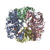
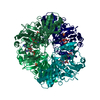
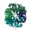
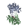
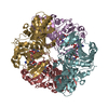
A: Glyceraldehyde-3-phosphate dehydrogenase, type I B: Glyceraldehyde-3-phosphate dehydrogenase, type I C: Glyceraldehyde-3-phosphate dehydrogenase, type I D: Glyceraldehyde-3-phosphate dehydrogenase, type I E: Glyceraldehyde-3-phosphate dehydrogenase, type I F: Glyceraldehyde-3-phosphate dehydrogenase, type I G: Glyceraldehyde-3-phosphate dehydrogenase, type I H: Glyceraldehyde-3-phosphate dehydrogenase, type I hetero molecules
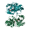
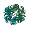
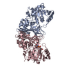
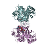
A: Glyceraldehyde-3-phosphate dehydrogenase, type I B: Glyceraldehyde-3-phosphate dehydrogenase, type I C: Glyceraldehyde-3-phosphate dehydrogenase, type I D: Glyceraldehyde-3-phosphate dehydrogenase, type I hetero molecules
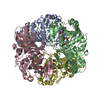
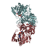
E: Glyceraldehyde-3-phosphate dehydrogenase, type I F: Glyceraldehyde-3-phosphate dehydrogenase, type I G: Glyceraldehyde-3-phosphate dehydrogenase, type I H: Glyceraldehyde-3-phosphate dehydrogenase, type I hetero molecules
Mass: 37194.086 Da / Num. of mol.: 8 Source method: isolated from a genetically manipulated source Details: expressed with a non-cleavable N-terminal hexahis tag Source: (gene. exp.) Burkholderia pseudomallei (bacteria) / Strain: 1710b / Gene: gap, BURPS1710b_3466 / Production host: Escherichia coli (E. coli) References: UniProt: Q3JNL6, Oxidoreductases; Acting on the aldehyde or oxo group of donors; With NAD+ or NADP+ as acceptor
Mass: 18.015 Da / Num. of mol.: 538 / Source method: isolated from a natural source / Formula: H2O
-
Experimental details
-
Experiment
Experiment
Method: X-RAY DIFFRACTION / Number of used crystals: 1
-
Sample preparation
Crystal
Density Matthews: 2.48 Å3/Da / Density % sol: 50.38 %
Crystal grow
Temperature: 289 K / Method: vapor diffusion, sitting drop Details: Molecular Dimensions PACT Premier screen condition E1, 25% PEG 3350, 0.2 M NaF with 25% glycerol as cryo-protectant, 33.4 mg/mL protein, crystal ID 201195e1, VAPOR DIFFUSION, SITTING DROP, temperature 289K
In the structure databanks used in Yorodumi, some data are registered as the other names, "COVID-19 virus" and "2019-nCoV". Here are the details of the virus and the list of structure data.
Jan 31, 2019. EMDB accession codes are about to change! (news from PDBe EMDB page)
EMDB accession codes are about to change! (news from PDBe EMDB page)
The allocation of 4 digits for EMDB accession codes will soon come to an end. Whilst these codes will remain in use, new EMDB accession codes will include an additional digit and will expand incrementally as the available range of codes is exhausted. The current 4-digit format prefixed with “EMD-” (i.e. EMD-XXXX) will advance to a 5-digit format (i.e. EMD-XXXXX), and so on. It is currently estimated that the 4-digit codes will be depleted around Spring 2019, at which point the 5-digit format will come into force.
The EM Navigator/Yorodumi systems omit the EMD- prefix.
Related info.:Q: What is EMD? / ID/Accession-code notation in Yorodumi/EM Navigator
Yorodumi is a browser for structure data from EMDB, PDB, SASBDB, etc.
This page is also the successor to EM Navigator detail page, and also detail information page/front-end page for Omokage search.
The word "yorodu" (or yorozu) is an old Japanese word meaning "ten thousand". "mi" (miru) is to see.
Related info.:EMDB / PDB / SASBDB / Comparison of 3 databanks / Yorodumi Search / Aug 31, 2016. New EM Navigator & Yorodumi / Yorodumi Papers / Jmol/JSmol / Function and homology information / Changes in new EM Navigator and Yorodumi
 Movie
Movie Controller

 Yorodumi
 Open data
 Basic information
 Components
 Glyceraldehyde 3-phosphate dehydrogenase
  Keywords
 Function and homology information
 Authors
 Citation
 Structure visualization
 Downloads & links
 Other downloads
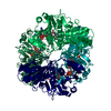


 PDBj
PDBj
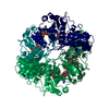
 Assembly
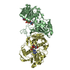

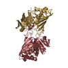

 Mass: 663.425 Da / Num. of mol.: 7 / Source method: obtained synthetically / Formula: C21H27N7O14P2 / Comment: NAD*YM
 Mass: 18.015 Da / Num. of mol.: 538 / Source method: isolated from a natural source / Formula: H2O
 Sample preparation
 / Beamline: 24-ID-C / Wavelength: 0.97934 Å
 Processing