 Movie
Movie Controller

[English] 日本語
 Yorodumi
- PDB-6leo: Crystal structure of thiosulfate transporter YeeE from Spirochaet... -
+ Open data

- Basic information
| Entry | Database: PDB / ID: 6leo | ||||||
|---|---|---|---|---|---|---|---|
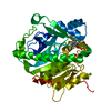
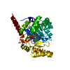
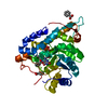
| Title | Crystal structure of thiosulfate transporter YeeE from Spirochaeta thermophila | ||||||
 Components Components | Sulf_transp domain-containing protein | ||||||
 Keywords Keywords |  TRANSPORT PROTEIN / Transmembrane / Transporter TRANSPORT PROTEIN / Transmembrane / Transporter | ||||||
| Function / homology | Sulphur transport domain / Sulphur transport / sulfate transport / plasma membrane / (2R)-2,3-dihydroxypropyl (9Z)-octadec-9-enoate / THIOSULFATE / Thiosulfate transporter TsuA Function and homology information Function and homology information | ||||||
| Biological species |   Spirochaeta thermophila (bacteria) Spirochaeta thermophila (bacteria) | ||||||
| Method | X-RAY DIFFRACTION / SYNCHROTRON / SIRAS / Resolution: 2.52 Å | ||||||
 Authors Authors | Tanaka, Y. / Tsukazaki, T. / Yoshikaie, K. / Takeuchi, A. / Uchino, S. / Sugano, Y. | ||||||
| Funding support |  Japan, 1items Japan, 1items
| ||||||
 Citation Citation | Journal: Sci Adv / Year: 2020 Title: Crystal structure of a YeeE/YedE family protein engaged in thiosulfate uptake. Authors: Tanaka, Y. / Yoshikaie, K. / Takeuchi, A. / Ichikawa, M. / Mori, T. / Uchino, S. / Sugano, Y. / Hakoshima, T. / Takagi, H. / Nonaka, G. / Tsukazaki, T. | ||||||
| History |
|
- Structure visualization
| Structure viewer | Molecule: MolmilJmol/JSmol |
|---|
- Downloads & links
-Download
| PDBx/mmCIF format | 6leo.cif.gz | 77.8 KB | Display | PDBx/mmCIF format |
|---|---|---|---|---|
| PDB format | pdb6leo.ent.gz | 62.6 KB | Display | PDB format |
| PDBx/mmJSON format | 6leo.json.gz | Tree view | PDBx/mmJSON format | |
| Others |  Other downloads Other downloads |
-Validation report
| Arichive directory | https://data.pdbj.org/pub/pdb/validation_reports/le/6leoftp://data.pdbj.org/pub/pdb/validation_reports/le/6leo | HTTPS FTP |
|---|
-Related structure data


-Links
 PDBj
PDBj


- Assembly
| Deposited unit | 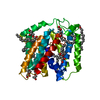
| ||||||||
|---|---|---|---|---|---|---|---|---|---|
| 1 |
| ||||||||
| Unit cell |
|
-Components
| #1: Protein | Mass: 36401.723 Da / Num. of mol.: 1 Source method: isolated from a genetically manipulated source Source: (gene. exp.) Spirochaeta thermophila (bacteria) / Gene: Spith_0734 / Plasmid: pET28a / Production host: Escherichia coli BL21(DE3) (bacteria) / Strain (production host): BL21(DE3) / Variant (production host): C41 / References: UniProt: G0GAP6 | ||||
|---|---|---|---|---|---|
| #2: Chemical | ChemComp-THJ / Thiosulfate  Mass: 112.128 Da / Num. of mol.: 1 / Source method: obtained synthetically / Formula: O3S2 / Feature type: SUBJECT OF INVESTIGATION Mass: 112.128 Da / Num. of mol.: 1 / Source method: obtained synthetically / Formula: O3S2 / Feature type: SUBJECT OF INVESTIGATION | ||||
| #3: Chemical | ChemComp-OLC / (   Mass: 356.540 Da / Num. of mol.: 9 / Source method: obtained synthetically / Formula: C21H40O4 Mass: 356.540 Da / Num. of mol.: 9 / Source method: obtained synthetically / Formula: C21H40O4#4: Water | ChemComp-HOH / | Water Mass: 18.015 Da / Num. of mol.: 23 / Source method: isolated from a natural source / Formula: H2O Mass: 18.015 Da / Num. of mol.: 23 / Source method: isolated from a natural source / Formula: H2OHas ligand of interest | Y | |
-Experimental details
-Experiment
| Experiment | Method: X-RAY DIFFRACTION / Number of used crystals: 1 |
|---|
- Sample preparation
| Crystal | Density Matthews: 2.44 Å3/Da / Density % sol: 49.59 % |
|---|---|
| Crystal grow | Temperature: 293 K / Method: lipidic cubic phase / pH: 7 / Details: Pentaerythritol-propoxylate, MES, NaCl / PH range: 6.5-7.5 |
-Data collection
| Diffraction | Mean temperature: 100 K / Serial crystal experiment: N | ||||||||||||||||||||||||||||||||||||||||||||||||||||||||||||||||||||||||||||||||||||||||||||||||||||
|---|---|---|---|---|---|---|---|---|---|---|---|---|---|---|---|---|---|---|---|---|---|---|---|---|---|---|---|---|---|---|---|---|---|---|---|---|---|---|---|---|---|---|---|---|---|---|---|---|---|---|---|---|---|---|---|---|---|---|---|---|---|---|---|---|---|---|---|---|---|---|---|---|---|---|---|---|---|---|---|---|---|---|---|---|---|---|---|---|---|---|---|---|---|---|---|---|---|---|---|---|---|
| Diffraction source | Source: SYNCHROTRON / Site: SPring-8 / Beamline: BL32XU / Wavelength: 1.44 Å | ||||||||||||||||||||||||||||||||||||||||||||||||||||||||||||||||||||||||||||||||||||||||||||||||||||
| Detector | Type: DECTRIS EIGER X 9M / Detector: PIXEL / Date: May 15, 2019 | ||||||||||||||||||||||||||||||||||||||||||||||||||||||||||||||||||||||||||||||||||||||||||||||||||||
| Radiation | Protocol: SINGLE WAVELENGTH / Monochromatic (M) / Laue (L): M / Scattering type: x-ray | ||||||||||||||||||||||||||||||||||||||||||||||||||||||||||||||||||||||||||||||||||||||||||||||||||||
| Radiation wavelength | Wavelength: 1.44 Å / Relative weight: 1 | ||||||||||||||||||||||||||||||||||||||||||||||||||||||||||||||||||||||||||||||||||||||||||||||||||||
| Reflection | Resolution: 2.52→43.134 Å / Num. obs: 12366 / % possible obs: 99.9 % / Redundancy: 87.621 % / Biso Wilson estimate: 45.497 Å2 / CC1/2: 0.984 / Rmerge(I) obs: 0.631 / Rrim(I) all: 0.635 / Χ2: 1.248 / Net I/σ(I): 14.76 / Num. measured all: 2039899 / Scaling rejects: 528 | ||||||||||||||||||||||||||||||||||||||||||||||||||||||||||||||||||||||||||||||||||||||||||||||||||||
| Reflection shell | Diffraction-ID: 1
|
- Processing
| Software |
| ||||||||||||||||||||||||||||||||||||||||||||||||||
|---|---|---|---|---|---|---|---|---|---|---|---|---|---|---|---|---|---|---|---|---|---|---|---|---|---|---|---|---|---|---|---|---|---|---|---|---|---|---|---|---|---|---|---|---|---|---|---|---|---|---|---|
| Refinement | Method to determine structure: SIRAS / Resolution: 2.52→43.134 Å / SU ML: 0.26 / Cross valid method: THROUGHOUT / σ(F): 1.34 / Phase error: 24.67
| ||||||||||||||||||||||||||||||||||||||||||||||||||
| Solvent computation | Shrinkage radii: 0.9 Å / VDW probe radii: 1.11 Å | ||||||||||||||||||||||||||||||||||||||||||||||||||
| Displacement parameters | Biso max: 109.78 Å2 / Biso mean: 36.597 Å2 / Biso min: 15.67 Å2 | ||||||||||||||||||||||||||||||||||||||||||||||||||
| Refinement step | Cycle: final / Resolution: 2.52→43.134 Å
| ||||||||||||||||||||||||||||||||||||||||||||||||||
| LS refinement shell | Refine-ID: X-RAY DIFFRACTION / Rfactor Rfree error: 0 / % reflection obs: 100 %
|
