 Movie
Movie Controller

[English] 日本語
 Yorodumi
- PDB-6ldt: K245A mutant of L-tyrosine decarboxylase from Methanocaldococcus ... -
+ Open data

- Basic information
| Entry | Database: PDB / ID: 6ldt | ||||||
|---|---|---|---|---|---|---|---|
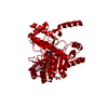
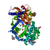
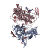
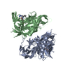
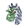
| Title | K245A mutant of L-tyrosine decarboxylase from Methanocaldococcus jannaschii complexed with a post-decarboxylation quinonoid-like intermediate formed with L-tyrosine | ||||||
 Components Components | L-tyrosine/L-aspartate decarboxylase | ||||||
 Keywords Keywords |  LYASE / PLP dependent decarboxylase / Catalytic Domain / Models / Molecular / Protein Conformation / PLP / Tyrosine / Quinonoid / Tyrosine Decarboxylase / Structure-Activity Relationship / Dunathan alignment LYASE / PLP dependent decarboxylase / Catalytic Domain / Models / Molecular / Protein Conformation / PLP / Tyrosine / Quinonoid / Tyrosine Decarboxylase / Structure-Activity Relationship / Dunathan alignment | ||||||
| Function / homology |  Function and homology information Function and homology informationmethanofuran biosynthetic process / tyrosine decarboxylase / tyrosine decarboxylase activity / aspartate 1-decarboxylase / aspartate 1-decarboxylase activity / coenzyme A biosynthetic process / carboxylic acid metabolic process / pyridoxal phosphate bindingSimilarity search - Function | ||||||
| Biological species |   Methanocaldococcus jannaschii (archaea) Methanocaldococcus jannaschii (archaea) | ||||||
| Method | X-RAY DIFFRACTION / MOLECULAR REPLACEMENT / Resolution: 1.93 Å | ||||||
 Authors Authors | Manoj, N. / Chellam Gayathri, S. | ||||||
 Citation Citation | Journal: J.Struct.Biol. / Year: 2019 Title: Structural insights into the mechanism of internal aldimine formation and catalytic loop dynamics in an archaeal Group II decarboxylase. Authors: Chellam Gayathri, S. / Manoj, N. #1: Journal: J. Struct. Biol. / Year: 2019Title: Structural insights into the mechanism of internal aldimine formation and catalytic loop dynamics in an archaeal Group II decarboxylase. Authors: Chellam Gayathri, S. / Manoj, N. | ||||||
| History |
|
- Structure visualization
| Structure viewer | Molecule: MolmilJmol/JSmol |
|---|
- Downloads & links
-Download
| PDBx/mmCIF format | 6ldt.cif.gz | 100.1 KB | Display | PDBx/mmCIF format |
|---|---|---|---|---|
| PDB format | pdb6ldt.ent.gz | 70.6 KB | Display | PDB format |
| PDBx/mmJSON format | 6ldt.json.gz | Tree view | PDBx/mmJSON format | |
| Others |  Other downloads Other downloads |
-Validation report
| Arichive directory | https://data.pdbj.org/pub/pdb/validation_reports/ld/6ldtftp://data.pdbj.org/pub/pdb/validation_reports/ld/6ldt | HTTPS FTP |
|---|
-Related structure data
| Related structure data | 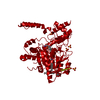 6jy1C  6ldrSC  6ldsC C: citing same article ( S: Starting model for refinement |
|---|---|
| Similar structure data |


-Links
 PDBj
PDBj- Assembly
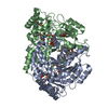
| Deposited unit | 
| ||||||||
|---|---|---|---|---|---|---|---|---|---|
| 1 | 
| ||||||||
| Unit cell |
| ||||||||
| Components on special symmetry positions |
|
-Components
| #1: Protein | Mass: 47600.730 Da / Num. of mol.: 1 / Mutation: K245A Source method: isolated from a genetically manipulated source Source: (gene. exp.) Methanocaldococcus jannaschii (strain ATCC 43067 / DSM 2661 / JAL-1 / JCM 10045 / NBRC 100440) (archaea)Strain: ATCC 43067 / DSM 2661 / JAL-1 / JCM 10045 / NBRC 100440 Gene: mfnA, MJ0050 / Production host:  Escherichia coli B (bacteria) / Strain (production host): B Escherichia coli B (bacteria) / Strain (production host): BReferences: UniProt: Q60358, aspartate 1-decarboxylase, tyrosine decarboxylase | ||||||||
|---|---|---|---|---|---|---|---|---|---|
| #2: Chemical | Glycerol  Mass: 92.094 Da / Num. of mol.: 3 / Source method: obtained synthetically / Formula: C3H8O3 Mass: 92.094 Da / Num. of mol.: 3 / Source method: obtained synthetically / Formula: C3H8O3#3: Chemical | ChemComp-SO4 / | Sulfate  Mass: 96.063 Da / Num. of mol.: 1 / Source method: obtained synthetically / Formula: SO4 Mass: 96.063 Da / Num. of mol.: 1 / Source method: obtained synthetically / Formula: SO4#4: Chemical | ChemComp-EBR / [( |   Mass: 366.306 Da / Num. of mol.: 1 / Source method: obtained synthetically / Formula: C16H19N2O6P / Feature type: SUBJECT OF INVESTIGATION Mass: 366.306 Da / Num. of mol.: 1 / Source method: obtained synthetically / Formula: C16H19N2O6P / Feature type: SUBJECT OF INVESTIGATION#5: Water | ChemComp-HOH / | Water Mass: 18.015 Da / Num. of mol.: 273 / Source method: isolated from a natural source / Formula: H2O Mass: 18.015 Da / Num. of mol.: 273 / Source method: isolated from a natural source / Formula: H2OHas ligand of interest | Y | |
-Experimental details
-Experiment
| Experiment | Method: X-RAY DIFFRACTION / Number of used crystals: 1 |
|---|
- Sample preparation
| Crystal | Density Matthews: 3.3 Å3/Da / Density % sol: 62.8 % |
|---|---|
| Crystal grow | Temperature: 293 K / Method: vapor diffusion, hanging drop / pH: 5.4 Details: 0.1M sodium citrate pH 5.4, 0.2M sodium potassium tartarate, 2.0M ammonium sulphate |
-Data collection
| Diffraction | Mean temperature: 100 K / Serial crystal experiment: N |
|---|---|
| Diffraction source | Source: ROTATING ANODE / Type: BRUKER AXS MICROSTAR / Wavelength: 1.5418 Å |
| Detector | Type: MARRESEARCH / Detector: IMAGE PLATE / Date: Mar 14, 2015 |
| Radiation | Monochromator: Double mirrors / Protocol: SINGLE WAVELENGTH / Monochromatic (M) / Laue (L): M / Scattering type: x-ray |
| Radiation wavelength | Wavelength: 1.5418 Å / Relative weight: 1 |
| Reflection | Resolution: 1.93→38.25 Å / Num. obs: 45548 / % possible obs: 100 % / Redundancy: 13 % / Biso Wilson estimate: 25.9 Å2 / CC1/2: 0.998 / Rmerge(I) obs: 0.129 / Rpim(I) all: 0.037 / Rrim(I) all: 0.134 / Net I/σ(I): 14.1 |
| Reflection shell | Resolution: 1.93→1.98 Å / Redundancy: 11.8 % / Rmerge(I) obs: 1.142 / Mean I/σ(I) obs: 2.3 / Num. unique obs: 2993 / CC1/2: 0.716 / Rpim(I) all: 0.342 / Rrim(I) all: 1.193 / % possible all: 99.8 |
- Processing
| Software |
| ||||||||||||||||||||||||
|---|---|---|---|---|---|---|---|---|---|---|---|---|---|---|---|---|---|---|---|---|---|---|---|---|---|
| Refinement | Method to determine structure: MOLECULAR REPLACEMENT Starting model: 6LDR Resolution: 1.93→38.25 Å / SU ML: 0.2 / Cross valid method: THROUGHOUT / σ(F): 1.33 / Phase error: 21.12
| ||||||||||||||||||||||||
| Solvent computation | Shrinkage radii: 0.9 Å / VDW probe radii: 1.11 Å | ||||||||||||||||||||||||
| Displacement parameters | Biso max: 65.29 Å2 / Biso mean: 27.8448 Å2 / Biso min: 10.82 Å2 | ||||||||||||||||||||||||
| Refinement step | Cycle: final / Resolution: 1.93→38.25 Å
| ||||||||||||||||||||||||
| Refine LS restraints |
| ||||||||||||||||||||||||
| LS refinement shell | Resolution: 1.93→1.97 Å / Rfactor Rfree error: 0
|
