 Movie
Movie Controller

[English] 日本語
 Yorodumi
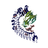
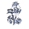
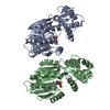
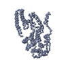
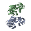
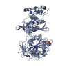
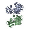
- PDB-6lb7: Crystal structure of the Ca2+-free and Ca2+-bound MICU1-MICU2 complex -
+ Open data

- Basic information
| Entry | Database: PDB / ID: 6lb7 | ||||||
|---|---|---|---|---|---|---|---|
| Title | Crystal structure of the Ca2+-free and Ca2+-bound MICU1-MICU2 complex | ||||||
 Components Components |
| ||||||
 Keywords Keywords | METAL BINDING PROTEIN /  Calcium binding protein / Mitochondrial / Uniporter / EF-hand Calcium binding protein / Mitochondrial / Uniporter / EF-hand | ||||||
| Function / homology |  Function and homology information Function and homology informationnegative regulation of mitochondrial calcium ion concentration / regulation of cellular hyperosmotic salinity response / Processing of SMDT1 / mitochondrial calcium ion transmembrane transport / uniplex complex / Mitochondrial calcium ion transport / calcium import into the mitochondrion / positive regulation of mitochondrial calcium ion concentration / mitochondrial calcium ion homeostasis / calcium ion import ...negative regulation of mitochondrial calcium ion concentration / regulation of cellular hyperosmotic salinity response / Processing of SMDT1 / mitochondrial calcium ion transmembrane transport / uniplex complex / Mitochondrial calcium ion transport / calcium import into the mitochondrion / positive regulation of mitochondrial calcium ion concentration / mitochondrial calcium ion homeostasis / calcium ion import / calcium channel complex / mitochondrial membrane / protein homooligomerization / mitochondrial intermembrane space / defense response / mitochondrial inner membrane / protein heterodimerization activity / calcium ion binding / mitochondrion / identical protein bindingSimilarity search - Function | ||||||
| Biological species |  Homo sapiens (human) Homo sapiens (human) | ||||||
| Method | X-RAY DIFFRACTION / SYNCHROTRON / MOLECULAR REPLACEMENT / Resolution: 2.101 Å | ||||||
 Authors Authors | Wu, W. / Shen, Q. / Zheng, J. / Jia, Z. | ||||||
| Funding support |  China, 1items China, 1items
| ||||||
 Citation Citation | |||||||
| History |
|
- Structure visualization
| Structure viewer | Molecule: MolmilJmol/JSmol |
|---|
- Downloads & links
-Download
| PDBx/mmCIF format | 6lb7.cif.gz | 526.9 KB | Display | PDBx/mmCIF format |
|---|---|---|---|---|
| PDB format | pdb6lb7.ent.gz | 429.2 KB | Display | PDB format |
| PDBx/mmJSON format | 6lb7.json.gz | Tree view | PDBx/mmJSON format | |
| Others |  Other downloads Other downloads |
-Validation report
| Arichive directory | https://data.pdbj.org/pub/pdb/validation_reports/lb/6lb7ftp://data.pdbj.org/pub/pdb/validation_reports/lb/6lb7 | HTTPS FTP |
|---|
-Related structure data
| Related structure data | 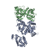 6lb8C 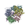 4nscS 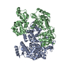 6iihS S: Starting model for refinement C: citing same article ( |
|---|---|
| Similar structure data |


-Links
 PDBj
PDBj

- Assembly
| Deposited unit | 
| ||||||||
|---|---|---|---|---|---|---|---|---|---|
| 1 | 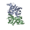
| ||||||||
| 2 | 
| ||||||||
| Unit cell |
|
-Components
| #1: Protein | Mass: 43083.953 Da / Num. of mol.: 2 Source method: isolated from a genetically manipulated source Source: (gene. exp.) Homo sapiens (human) / Gene: MICU1, CALC, CBARA1 / Plasmid: pET-28b(+) / Production host:  Escherichia coli BL21(DE3) (bacteria) / Strain (production host): BL21(DE3) / References: UniProt: Q9BPX6 Escherichia coli BL21(DE3) (bacteria) / Strain (production host): BL21(DE3) / References: UniProt: Q9BPX6#2: Protein | Mass: 38719.238 Da / Num. of mol.: 2 Source method: isolated from a genetically manipulated source Source: (gene. exp.) Homo sapiens (human) / Gene: MICU2, EFHA1 / Plasmid: pET-28b(+) / Production host: Escherichia coli BL21(DE3) (bacteria) / Strain (production host): BL21(DE3) / References: UniProt: Q8IYU8#3: Chemical |   Mass: 40.078 Da / Num. of mol.: 2 / Source method: obtained synthetically / Formula: Ca / Feature type: SUBJECT OF INVESTIGATION Mass: 40.078 Da / Num. of mol.: 2 / Source method: obtained synthetically / Formula: Ca / Feature type: SUBJECT OF INVESTIGATION#4: Water | ChemComp-HOH / | Water Mass: 18.015 Da / Num. of mol.: 1098 / Source method: isolated from a natural source / Formula: H2O Mass: 18.015 Da / Num. of mol.: 1098 / Source method: isolated from a natural source / Formula: H2OHas ligand of interest | Y | |
|---|
-Experimental details
-Experiment
| Experiment | Method: X-RAY DIFFRACTION / Number of used crystals: 1 |
|---|
- Sample preparation
| Crystal | Density Matthews: 2.54 Å3/Da / Density % sol: 51.6 % |
|---|---|
| Crystal grow | Temperature: 293.15 K / Method: vapor diffusion, hanging drop / pH: 5 Details: 0.2 M ammonium chloride, 5% (w/v) polyethylene glycol (PEG) 3350 |
-Data collection
| Diffraction | Mean temperature: 100 K / Serial crystal experiment: N |
|---|---|
| Diffraction source | Source: SYNCHROTRON / Site: SSRF / Beamline: BL19U1 / Wavelength: 0.97892 Å |
| Detector | Type: DECTRIS PILATUS3 6M / Detector: PIXEL / Date: Oct 3, 2018 |
| Radiation | Protocol: SINGLE WAVELENGTH / Monochromatic (M) / Laue (L): M / Scattering type: x-ray |
| Radiation wavelength | Wavelength: 0.97892 Å / Relative weight: 1 |
| Reflection | Resolution: 2.101→47.3 Å / Num. obs: 92168 / % possible obs: 98 % / Redundancy: 13 % / CC1/2: 0.984 / Rmerge(I) obs: 0.1246 / Rrim(I) all: 0.1297 / Net I/σ(I): 10.87 |
| Reflection shell | Resolution: 2.101→2.176 Å / Redundancy: 12.3 % / Rmerge(I) obs: 1.192 / Mean I/σ(I) obs: 1.35 / Num. unique obs: 8058 / CC1/2: 0.775 / % possible all: 100 |
- Processing
| Software |
| ||||||||||||||||||||||||||||||||||||||||||||||||||||||||||||||||||||||||||||||||||||||||||
|---|---|---|---|---|---|---|---|---|---|---|---|---|---|---|---|---|---|---|---|---|---|---|---|---|---|---|---|---|---|---|---|---|---|---|---|---|---|---|---|---|---|---|---|---|---|---|---|---|---|---|---|---|---|---|---|---|---|---|---|---|---|---|---|---|---|---|---|---|---|---|---|---|---|---|---|---|---|---|---|---|---|---|---|---|---|---|---|---|---|---|---|
| Refinement | Method to determine structure: MOLECULAR REPLACEMENT Starting model: 4NSC, 6IIH Resolution: 2.101→47.3 Å / SU ML: 0.2 / Cross valid method: FREE R-VALUE / σ(F): 1.34 / Phase error: 22.45
| ||||||||||||||||||||||||||||||||||||||||||||||||||||||||||||||||||||||||||||||||||||||||||
| Solvent computation | Shrinkage radii: 0.9 Å / VDW probe radii: 1.11 Å | ||||||||||||||||||||||||||||||||||||||||||||||||||||||||||||||||||||||||||||||||||||||||||
| Displacement parameters | Biso max: 129.94 Å2 / Biso mean: 35.5483 Å2 / Biso min: 9.42 Å2 | ||||||||||||||||||||||||||||||||||||||||||||||||||||||||||||||||||||||||||||||||||||||||||
| Refinement step | Cycle: final / Resolution: 2.101→47.3 Å
| ||||||||||||||||||||||||||||||||||||||||||||||||||||||||||||||||||||||||||||||||||||||||||
| Refine LS restraints |
| ||||||||||||||||||||||||||||||||||||||||||||||||||||||||||||||||||||||||||||||||||||||||||
| LS refinement shell | Refine-ID: X-RAY DIFFRACTION / Rfactor Rfree error: 0
|
