 Movie
Movie Controller


+ Open data

- Basic information
| Entry | Database: PDB / ID: 6kx0 | |||||||||
|---|---|---|---|---|---|---|---|---|---|---|
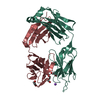
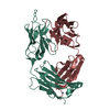
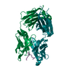
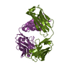
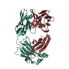
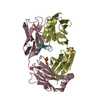
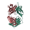
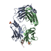
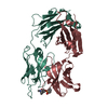
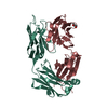
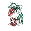
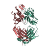
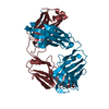
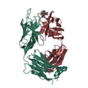
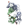
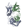
| Title | Crystal structure of SN-101 mAb non-liganded form | |||||||||
 Components Components |
| |||||||||
 Keywords Keywords | IMMUNE SYSTEM / Antibody / IgG1 / MUC1 / Glycopeptide | |||||||||
| Biological species |  | |||||||||
| Method |  X-RAY DIFFRACTION / SYNCHROTRON / MOLECULAR REPLACEMENT / Resolution: 2.404 Å X-RAY DIFFRACTION / SYNCHROTRON / MOLECULAR REPLACEMENT / Resolution: 2.404 Å | |||||||||
 Authors Authors | Wakui, H. / Tanaka, Y. / Kato, K. / Ose, T. / Matsumoto, I. / Min, Y. / Tachibana, T. / Nishimura, S.-I. | |||||||||
| Funding support |  Japan, 2items Japan, 2items
| |||||||||
 Citation Citation | Journal: Chem Sci / Year: 2020 Title: A straightforward approach to antibodies recognising cancer specific glycopeptidic neoepitopes Authors: Wakui, H. / Tanaka, Y. / Ose, T. / Matsumoto, I. / Kato, K. / Min, Y. / Tachibana, T. / Sato, M. / Naruchi, K. / Martin, F.G. / Hinou, H. / Nishimura, S.-I. | |||||||||
| History |
|
- Structure visualization
| Structure viewer | Molecule:  MolmilJmol/JSmol MolmilJmol/JSmol |
|---|
- Downloads & links
-Download
| PDBx/mmCIF format | 6kx0.cif.gz | 100.4 KB | Display | PDBx/mmCIF format |
|---|---|---|---|---|
| PDB format | pdb6kx0.ent.gz | 72.5 KB | Display | PDB format |
| PDBx/mmJSON format | 6kx0.json.gz | Tree view | PDBx/mmJSON format | |
| Others |  Other downloads Other downloads |
-Validation report
| Summary document | 6kx0_validation.pdf.gz | 458.5 KB | Display | wwPDB validaton report |
|---|---|---|---|---|
| Full document | 6kx0_full_validation.pdf.gz | 463.8 KB | Display | |
| Data in XML | 6kx0_validation.xml.gz | 17.5 KB | Display | |
| Data in CIF | 6kx0_validation.cif.gz | 23.2 KB | Display | |
| Arichive directory | https://data.pdbj.org/pub/pdb/validation_reports/kx/6kx0ftp://data.pdbj.org/pub/pdb/validation_reports/kx/6kx0 | HTTPS FTP |
-Related structure data
| Related structure data |  6kx1C 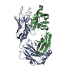 1pz5S S: Starting model for refinement C: citing same article ( |
|---|---|
| Similar structure data |


-Links
 PDBj
PDBj

- Assembly
| Deposited unit | 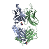
| ||||||||||||
|---|---|---|---|---|---|---|---|---|---|---|---|---|---|
| 1 |
| ||||||||||||
| Unit cell |
|
-Components
| #1: Antibody | Mass: 25541.768 Da / Num. of mol.: 1 Source method: isolated from a genetically manipulated source Source: (gene. exp.) | ||||
|---|---|---|---|---|---|
| #2: Antibody | Mass: 26438.670 Da / Num. of mol.: 1 Source method: isolated from a genetically manipulated source Source: (gene. exp.) | ||||
| #3: Chemical |   Mass: 122.143 Da / Num. of mol.: 2 / Source method: obtained synthetically / Formula: C4H12NO3 / Comment: pH buffer*YM Mass: 122.143 Da / Num. of mol.: 2 / Source method: obtained synthetically / Formula: C4H12NO3 / Comment: pH buffer*YM#4: Water | ChemComp-HOH / |  Mass: 18.015 Da / Num. of mol.: 37 / Source method: isolated from a natural source / Formula: H2O Mass: 18.015 Da / Num. of mol.: 37 / Source method: isolated from a natural source / Formula: H2OHas ligand of interest | N | |
-Experimental details
-Experiment
| Experiment | Method: X-RAY DIFFRACTION / Number of used crystals: 1 |
|---|
- Sample preparation
| Crystal | Density Matthews: 2.06 Å3/Da / Density % sol: 40.26 % |
|---|---|
| Crystal grow | Temperature: 293 K / Method: vapor diffusion, sitting drop / pH: 8 / Details: 300-350 mM NaSCN 20-30% PEG3350 |
-Data collection
| Diffraction | Mean temperature: 100 K / Serial crystal experiment: N |
|---|---|
| Diffraction source | Source: SYNCHROTRON / Site: Photon Factory / Beamline: BL-17A / Wavelength: 0.98 Å |
| Detector | Type: DECTRIS PILATUS 2M / Detector: PIXEL / Date: Mar 5, 2017 |
| Radiation | Protocol: SINGLE WAVELENGTH / Monochromatic (M) / Laue (L): M / Scattering type: x-ray |
| Radiation wavelength | Wavelength: 0.98 Å / Relative weight: 1 |
| Reflection | Resolution: 2.4→50 Å / Num. obs: 16818 / % possible obs: 99.6 % / Redundancy: 4.99 % / Rrim(I) all: 0.101 / Net I/σ(I): 12.78 |
| Reflection shell | Resolution: 2.4→2.55 Å / Redundancy: 5.08 % / Num. unique obs: 2665 / Rrim(I) all: 0.663 / % possible all: 98.9 |
- Processing
| Software |
| ||||||||||||||||||||||||
|---|---|---|---|---|---|---|---|---|---|---|---|---|---|---|---|---|---|---|---|---|---|---|---|---|---|
| Refinement | Method to determine structure: MOLECULAR REPLACEMENT Starting model: 1PZ5 Resolution: 2.404→39.378 Å / Cross valid method: FREE R-VALUE
| ||||||||||||||||||||||||
| Displacement parameters | Biso mean: 51.59 Å2 | ||||||||||||||||||||||||
| Refinement step | Cycle: LAST / Resolution: 2.404→39.378 Å
| ||||||||||||||||||||||||
| Refine LS restraints |
|
