 Movie
Movie Controller

[English] 日本語
 Yorodumi
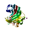
- PDB-6kwf: Crystal Structure Analysis of Endo-beta-1,4-xylanase II Complexed... -
+ Open data

- Basic information
| Entry | Database: PDB / ID: 6kwf | ||||||
|---|---|---|---|---|---|---|---|
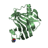
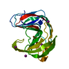
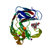
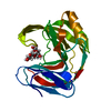
| Title | Crystal Structure Analysis of Endo-beta-1,4-xylanase II Complexed with Xylotriose | ||||||
 Components Components | Endo-1,4-beta-xylanase 2 Xylanase Xylanase | ||||||
 Keywords Keywords | HYDROLASE / Xylanase II / Xylotriose | ||||||
| Function / homology |  Function and homology informationendo-1,4-beta-xylanase activity / endo-1,4-beta-xylanase / xylan catabolic process / extracellular region Function and homology informationendo-1,4-beta-xylanase activity / endo-1,4-beta-xylanase / xylan catabolic process / extracellular regionSimilarity search - Function | ||||||
| Biological species |  Trichoderma reesei RUT C-30 (fungus) Trichoderma reesei RUT C-30 (fungus) | ||||||
| Method | X-RAY DIFFRACTION / SYNCHROTRON / MOLECULAR REPLACEMENT / Resolution: 1.22 Å | ||||||
 Authors Authors | Li, C. / Wan, Q. | ||||||
| Funding support |  China, 1items China, 1items
| ||||||
 Citation Citation | Journal: Protein J. / Year: 2020 Title: Studying the Role of a Single Mutation of a Family 11 Glycoside Hydrolase Using High-Resolution X-ray Crystallography. Authors: Li, Z. / Zhang, X. / Li, C. / Kovalevsky, A. / Wan, Q. | ||||||
| History |
|
- Structure visualization
| Structure viewer | Molecule: MolmilJmol/JSmol |
|---|
- Downloads & links
-Download
| PDBx/mmCIF format | 6kwf.cif.gz | 95.6 KB | Display | PDBx/mmCIF format |
|---|---|---|---|---|
| PDB format | pdb6kwf.ent.gz | 72.1 KB | Display | PDB format |
| PDBx/mmJSON format | 6kwf.json.gz | Tree view | PDBx/mmJSON format | |
| Others |  Other downloads Other downloads |
-Validation report
| Arichive directory | https://data.pdbj.org/pub/pdb/validation_reports/kw/6kwfftp://data.pdbj.org/pub/pdb/validation_reports/kw/6kwf | HTTPS FTP |
|---|
-Related structure data
| Related structure data |  6jugC 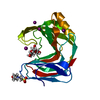 6jwbC  6k9oC  6k9rC  6k9wC  6kw9C  6kwcC 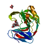 6kwdC 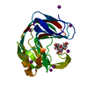 6kwgC  2dfcS S: Starting model for refinement C: citing same article ( |
|---|---|
| Similar structure data |


-Links
 PDBj
PDBj
- Assembly
| Deposited unit | 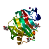
| |||||||||
|---|---|---|---|---|---|---|---|---|---|---|
| 1 |
| |||||||||
| Unit cell |
| |||||||||
| Components on special symmetry positions |
|
-Components
-Protein , 1 types, 1 molecules A
| #1: Protein | Xylanase / Xylanase 2 / 1 / 4-beta-D-xylan xylanohydrolase 2 / Alkaline endo-beta-1 / 4-xylanase Mass: 20815.400 Da / Num. of mol.: 1 / Mutation: N44D Source method: isolated from a genetically manipulated source Source: (gene. exp.) Trichoderma reesei RUT C-30 (fungus) / Gene: xyn2, M419DRAFT_124931 / Production host:  Escherichia coli (E. coli) / References: UniProt: P36217, endo-1,4-beta-xylanase Escherichia coli (E. coli) / References: UniProt: P36217, endo-1,4-beta-xylanase |
|---|
-Sugars , 2 types, 2 molecules 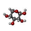
| #2: Polysaccharide | beta-D-xylopyranose-(1-4)-beta-D-xylopyranose-(1-4)-beta-D-xylopyranose / 4beta-beta-xylotriose  , Oligosaccharide / Class: Metabolism / Mass: 414.360 Da / Num. of mol.: 1 , Oligosaccharide / Class: Metabolism / Mass: 414.360 Da / Num. of mol.: 1Source method: isolated from a genetically manipulated source Details: oligosaccharide / References: 4beta-beta-xylotriose |
|---|---|
| #3: Sugar | ChemComp-XYP / Xylose Type: D-saccharide, beta linking / Mass: 150.130 Da / Num. of mol.: 1 Type: D-saccharide, beta linking / Mass: 150.130 Da / Num. of mol.: 1Source method: isolated from a genetically manipulated source Formula: C5H10O5 / Feature type: SUBJECT OF INVESTIGATION |
-Non-polymers , 3 types, 277 molecules 


| #4: Chemical | Glycerol Mass: 92.094 Da / Num. of mol.: 2 / Source method: obtained synthetically / Formula: C3H8O3 / Feature type: SUBJECT OF INVESTIGATION Mass: 92.094 Da / Num. of mol.: 2 / Source method: obtained synthetically / Formula: C3H8O3 / Feature type: SUBJECT OF INVESTIGATION#5: Chemical | ChemComp-IOD / Iodide Mass: 126.904 Da / Num. of mol.: 4 / Source method: obtained synthetically / Formula: I / Feature type: SUBJECT OF INVESTIGATION Mass: 126.904 Da / Num. of mol.: 4 / Source method: obtained synthetically / Formula: I / Feature type: SUBJECT OF INVESTIGATION#6: Water | ChemComp-HOH / | WaterMass: 18.015 Da / Num. of mol.: 271 / Source method: isolated from a natural source / Formula: H2O |
|---|
-Details
| Has ligand of interest | Y |
|---|
-Experimental details
-Experiment
| Experiment | Method: X-RAY DIFFRACTION / Number of used crystals: 1 |
|---|
- Sample preparation
| Crystal | Density Matthews: 2.14 Å3/Da / Density % sol: 42.62 % |
|---|---|
| Crystal grow | Temperature: 291 K / Method: evaporation / pH: 4.5 / Details: PEG 8000, NaI, NaAc-HAc |
-Data collection
| Diffraction | Mean temperature: 80 K / Serial crystal experiment: N |
|---|---|
| Diffraction source | Source: SYNCHROTRON / Site: SSRF / Beamline: BL18U1 / Wavelength: 1 Å |
| Detector | Type: ADSC QUANTUM 315r / Detector: CCD / Date: Jun 2, 2019 |
| Radiation | Protocol: SINGLE WAVELENGTH / Monochromatic (M) / Laue (L): M / Scattering type: x-ray |
| Radiation wavelength | Wavelength: 1 Å / Relative weight: 1 |
| Reflection | Resolution: 1.22→19.84 Å / Num. obs: 52630 / % possible obs: 98.3 % / Redundancy: 1.6 % / Biso Wilson estimate: 11.68 Å2 / Rmerge(I) obs: 0.91 / Net I/σ(I): 8.24 |
| Reflection shell | Resolution: 1.22→1.264 Å / Rmerge(I) obs: 0.91 / Num. unique obs: 5303 |
- Processing
| Software |
| ||||||||||||||||||||||||||||||||||||||||||||||||||||||||||||||||||
|---|---|---|---|---|---|---|---|---|---|---|---|---|---|---|---|---|---|---|---|---|---|---|---|---|---|---|---|---|---|---|---|---|---|---|---|---|---|---|---|---|---|---|---|---|---|---|---|---|---|---|---|---|---|---|---|---|---|---|---|---|---|---|---|---|---|---|---|
| Refinement | Method to determine structure: MOLECULAR REPLACEMENT Starting model: 2DFC Resolution: 1.22→19.839 Å / SU ML: 0.1 / Cross valid method: THROUGHOUT / σ(F): 1.71 / Phase error: 15.19
| ||||||||||||||||||||||||||||||||||||||||||||||||||||||||||||||||||
| Solvent computation | Shrinkage radii: 0.9 Å / VDW probe radii: 1.11 Å | ||||||||||||||||||||||||||||||||||||||||||||||||||||||||||||||||||
| Displacement parameters | Biso max: 91.52 Å2 / Biso mean: 15.7247 Å2 / Biso min: 4.98 Å2 | ||||||||||||||||||||||||||||||||||||||||||||||||||||||||||||||||||
| Refinement step | Cycle: final / Resolution: 1.22→19.839 Å
| ||||||||||||||||||||||||||||||||||||||||||||||||||||||||||||||||||
| Refine LS restraints |
| ||||||||||||||||||||||||||||||||||||||||||||||||||||||||||||||||||
| LS refinement shell | Refine-ID: X-RAY DIFFRACTION / Rfactor Rfree error: 0
|
