 Movie
Movie Controller


+ Open data

- Basic information
| Entry | Database: PDB / ID: 6ksv | ||||||
|---|---|---|---|---|---|---|---|
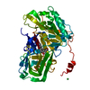
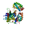
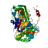
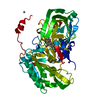
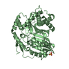
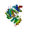
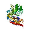
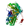
| Title | Crystal structure of SurE with D-Leu | ||||||
 Components Components | Alpha/beta hydrolase Alpha/beta hydrolase superfamily Alpha/beta hydrolase superfamily | ||||||
 Keywords Keywords | HYDROLASE / NRPS / cyclase / PBP-binding protein | ||||||
| Function / homology |  Function and homology information Function and homology information | ||||||
| Biological species |  Streptomyces albidoflavus (bacteria) Streptomyces albidoflavus (bacteria) | ||||||
| Method | X-RAY DIFFRACTION / SYNCHROTRON / MOLECULAR REPLACEMENT / Resolution: 2.42 Å | ||||||
 Authors Authors | Zhai, R. / Mori, T. / Abe, I. | ||||||
 Citation Citation | Journal: Nat Catal / Year: 2020 Title: Heterochiral coupling in non-ribosomal peptide macrolactamization Authors: Matsuda, K. / Zhai, R. / Mori, T. / Kobayashi, M. / Sano, A. / Abe, I. / Wakimoto, T. | ||||||
| History |
|
- Structure visualization
| Structure viewer | Molecule: MolmilJmol/JSmol |
|---|
- Downloads & links
-Download
| PDBx/mmCIF format | 6ksv.cif.gz | 175.5 KB | Display | PDBx/mmCIF format |
|---|---|---|---|---|
| PDB format | pdb6ksv.ent.gz | 135.5 KB | Display | PDB format |
| PDBx/mmJSON format | 6ksv.json.gz | Tree view | PDBx/mmJSON format | |
| Others |  Other downloads Other downloads |
-Validation report
| Arichive directory | https://data.pdbj.org/pub/pdb/validation_reports/ks/6ksvftp://data.pdbj.org/pub/pdb/validation_reports/ks/6ksv | HTTPS FTP |
|---|
-Related structure data
| Related structure data | 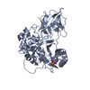 6ksuSC S: Starting model for refinement C: citing same article ( |
|---|---|
| Similar structure data |

-Links
 PDBj
PDBj


- Assembly
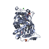
| Deposited unit | 
| ||||||||
|---|---|---|---|---|---|---|---|---|---|
| 1 | 
| ||||||||
| 2 | 
| ||||||||
| Unit cell |
|
-Components
-Protein , 1 types, 2 molecules AB
| #1: Protein | Alpha/beta hydrolase superfamily / SurE Mass: 49642.664 Da / Num. of mol.: 2 Source method: isolated from a genetically manipulated source Source: (gene. exp.) Streptomyces albidoflavus (bacteria) / Plasmid: pET28a / Production host: Escherichia coli BL21(DE3) (bacteria) / References: UniProt: A0A4Q6RSJ1, UniProt: A0A679G4U8*PLUS |
|---|
-Non-polymers , 5 types, 118 molecules 


| #2: Chemical | ChemComp-DLE / Leucine Type: D-peptide linking / Mass: 131.173 Da / Num. of mol.: 1 / Source method: obtained synthetically / Formula: C6H13NO2 / Feature type: SUBJECT OF INVESTIGATION Type: D-peptide linking / Mass: 131.173 Da / Num. of mol.: 1 / Source method: obtained synthetically / Formula: C6H13NO2 / Feature type: SUBJECT OF INVESTIGATION | ||||||
|---|---|---|---|---|---|---|---|
| #3: Chemical | ChemComp-SO4 / Sulfate Mass: 96.063 Da / Num. of mol.: 5 / Source method: obtained synthetically / Formula: SO4 Mass: 96.063 Da / Num. of mol.: 5 / Source method: obtained synthetically / Formula: SO4#4: Chemical | Yttrium Mass: 88.906 Da / Num. of mol.: 3 / Source method: obtained synthetically / Formula: Y Mass: 88.906 Da / Num. of mol.: 3 / Source method: obtained synthetically / Formula: Y#5: Chemical | ChemComp-E7L / |  Mass: 232.343 Da / Num. of mol.: 1 / Source method: obtained synthetically / Formula: C10H20N2O2S / Feature type: SUBJECT OF INVESTIGATION Mass: 232.343 Da / Num. of mol.: 1 / Source method: obtained synthetically / Formula: C10H20N2O2S / Feature type: SUBJECT OF INVESTIGATION#6: Water | ChemComp-HOH / | WaterMass: 18.015 Da / Num. of mol.: 108 / Source method: isolated from a natural source / Formula: H2O |
-Details
| Has ligand of interest | Y |
|---|
-Experimental details
-Experiment
| Experiment | Method: X-RAY DIFFRACTION / Number of used crystals: 1 |
|---|
- Sample preparation
| Crystal | Density Matthews: 2.2 Å3/Da / Density % sol: 44.07 % |
|---|---|
| Crystal grow | Temperature: 277 K / Method: vapor diffusion, sitting drop / pH: 7.5 / Details: ammonium sulfate, Yttrium Chloride, TCEP |
-Data collection
| Diffraction | Mean temperature: 277 K / Serial crystal experiment: N |
|---|---|
| Diffraction source | Source: SYNCHROTRON / Site: Photon Factory  / Beamline: BL-1A / Wavelength: 1 Å / Beamline: BL-1A / Wavelength: 1 Å |
| Detector | Type: DECTRIS EIGER X 4M / Detector: PIXEL / Date: Feb 18, 2019 |
| Radiation | Protocol: SINGLE WAVELENGTH / Monochromatic (M) / Laue (L): M / Scattering type: x-ray |
| Radiation wavelength | Wavelength: 1 Å / Relative weight: 1 |
| Reflection | Resolution: 2.4197→50 Å / Num. obs: 32476 / % possible obs: 99.2 % / Redundancy: 3.5 % / Biso Wilson estimate: 44.23 Å2 / CC1/2: 0.997 / Rmerge(I) obs: 0.076 / Net I/σ(I): 11.41 |
| Reflection shell | Resolution: 2.4197→2.57 Å / Redundancy: 3.5 % / Rmerge(I) obs: 0.507 / Mean I/σ(I) obs: 2.44 / Num. unique obs: 5134 / CC1/2: 0.839 / % possible all: 97.7 |
- Processing
| Software |
| ||||||||||||||||||||||||||||||||||||||||||||||||||||||||||||||||||||||||||||||
|---|---|---|---|---|---|---|---|---|---|---|---|---|---|---|---|---|---|---|---|---|---|---|---|---|---|---|---|---|---|---|---|---|---|---|---|---|---|---|---|---|---|---|---|---|---|---|---|---|---|---|---|---|---|---|---|---|---|---|---|---|---|---|---|---|---|---|---|---|---|---|---|---|---|---|---|---|---|---|---|
| Refinement | Method to determine structure: MOLECULAR REPLACEMENT Starting model: 6KSU Resolution: 2.42→45.086 Å / SU ML: 0.37 / Cross valid method: THROUGHOUT / σ(F): 1.38 / Phase error: 30.23
| ||||||||||||||||||||||||||||||||||||||||||||||||||||||||||||||||||||||||||||||
| Solvent computation | Shrinkage radii: 0.9 Å / VDW probe radii: 1.11 Å | ||||||||||||||||||||||||||||||||||||||||||||||||||||||||||||||||||||||||||||||
| Displacement parameters | Biso max: 92.27 Å2 / Biso mean: 43.9924 Å2 / Biso min: 17.47 Å2 | ||||||||||||||||||||||||||||||||||||||||||||||||||||||||||||||||||||||||||||||
| Refinement step | Cycle: final / Resolution: 2.42→45.086 Å
| ||||||||||||||||||||||||||||||||||||||||||||||||||||||||||||||||||||||||||||||
| LS refinement shell | Refine-ID: X-RAY DIFFRACTION / Rfactor Rfree error: 0
|
