 Movie
Movie Controller

[English] 日本語
 Yorodumi
- PDB-5t3e: Crystal structure of a nonribosomal peptide synthetase heterocycl... -
+ Open data

- Basic information
| Entry | Database: PDB / ID: 5t3e | ||||||
|---|---|---|---|---|---|---|---|
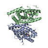
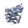
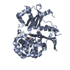
| Title | Crystal structure of a nonribosomal peptide synthetase heterocyclization domain. | ||||||
 Components Components | Bacillamide synthetase heterocyclization domain | ||||||
 Keywords Keywords |  LIGASE / nonribosomal peptide synthetase / heterocyclization domain / natural products / thiazoline LIGASE / nonribosomal peptide synthetase / heterocyclization domain / natural products / thiazoline | ||||||
| Function / homology |  Function and homology information Function and homology information | ||||||
| Biological species |  Thermoactinomyces vulgaris (bacteria) Thermoactinomyces vulgaris (bacteria) | ||||||
| Method | X-RAY DIFFRACTION / SYNCHROTRON / MOLECULAR REPLACEMENT / Resolution: 2.297 Å | ||||||
 Authors Authors | Bloudoff, K. / Schmeing, T.M. | ||||||
 Citation Citation | Journal: Proc. Natl. Acad. Sci. U.S.A. / Year: 2017 Title: Structural and mutational analysis of the nonribosomal peptide synthetase heterocyclization domain provides insight into catalysis. Authors: Bloudoff, K. / Fage, C.D. / Marahiel, M.A. / Schmeing, T.M. | ||||||
| History |
|
- Structure visualization
| Structure viewer | Molecule: MolmilJmol/JSmol |
|---|
- Downloads & links
-Download
| PDBx/mmCIF format | 5t3e.cif.gz | 188.1 KB | Display | PDBx/mmCIF format |
|---|---|---|---|---|
| PDB format | pdb5t3e.ent.gz | 149.3 KB | Display | PDB format |
| PDBx/mmJSON format | 5t3e.json.gz | Tree view | PDBx/mmJSON format | |
| Others |  Other downloads Other downloads |
-Validation report
| Arichive directory | https://data.pdbj.org/pub/pdb/validation_reports/t3/5t3eftp://data.pdbj.org/pub/pdb/validation_reports/t3/5t3e | HTTPS FTP |
|---|
-Related structure data
| Related structure data | 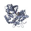 4jn3S S: Starting model for refinement |
|---|---|
| Similar structure data |


-Links
 PDBj
PDBj


- Assembly
| Deposited unit | 
| ||||||||
|---|---|---|---|---|---|---|---|---|---|
| 1 | 
| ||||||||
| 2 | 
| ||||||||
| 3 |
| ||||||||
| Unit cell |
|
-Components
| #1: Protein | Mass: 51775.801 Da / Num. of mol.: 2 / Fragment: unp residues 844-1287 Source method: isolated from a genetically manipulated source Source: (gene. exp.) Thermoactinomyces vulgaris (bacteria) / Gene: ADL26_17380 / Production host: Escherichia coli (E. coli) / References: UniProt: A0A0N0Y601#2: Chemical | Sulfate  Mass: 96.063 Da / Num. of mol.: 2 / Source method: obtained synthetically / Formula: SO4 Mass: 96.063 Da / Num. of mol.: 2 / Source method: obtained synthetically / Formula: SO4#3: Water | ChemComp-HOH / | Water Mass: 18.015 Da / Num. of mol.: 217 / Source method: isolated from a natural source / Formula: H2O Mass: 18.015 Da / Num. of mol.: 217 / Source method: isolated from a natural source / Formula: H2O |
|---|
-Experimental details
-Experiment
| Experiment | Method: X-RAY DIFFRACTION / Number of used crystals: 1 |
|---|
- Sample preparation
| Crystal | Density Matthews: 2.89 Å3/Da / Density % sol: 57.46 % |
|---|---|
| Crystal grow | Temperature: 295 K / Method: vapor diffusion, sitting drop Details: BmdB-Cy2 was crystallized by mixing with 0.88% Tween 20, 1.62 M ammonium sulfate, 0.1 M HEPES pH 7.5, 2.67% PEG 400, and 3% 6-aminohexanoic acid, and equilibrated 0.88% Tween 20, 1.62 M ...Details: BmdB-Cy2 was crystallized by mixing with 0.88% Tween 20, 1.62 M ammonium sulfate, 0.1 M HEPES pH 7.5, 2.67% PEG 400, and 3% 6-aminohexanoic acid, and equilibrated 0.88% Tween 20, 1.62 M ammonium sulfate, 0.1 M HEPES pH 7.5, 2.67% PEG 400, and 3% 6-aminohexanoic acid. Crystals were transferred into a solution of mother liquor plus 20% (v/v) ethylene glycol, and flash-cooled in liquid nitrogen. |
-Data collection
| Diffraction | Mean temperature: 100 K |
|---|---|
| Diffraction source | Source: SYNCHROTRON / Site: CLSI  / Beamline: 08ID-1 / Wavelength: 0.97949 Å / Beamline: 08ID-1 / Wavelength: 0.97949 Å |
| Detector | Type: RAYONIX MX-300 / Detector: CCD / Date: Oct 7, 2015 |
| Radiation | Protocol: SINGLE WAVELENGTH / Monochromatic (M) / Laue (L): M / Scattering type: x-ray |
| Radiation wavelength | Wavelength: 0.97949 Å / Relative weight: 1 |
| Reflection | Resolution: 2.3→39.9 Å / Num. obs: 52064 / % possible obs: 99.8 % / Redundancy: 3.1 % / Net I/σ(I): 15.1 |
- Processing
| Software |
| ||||||||||||||||||||||||||||||||||||||||||||||||||||||||||||||||||||||||||||||||||||||||||||||||||||||||||||||||||||||||||||||||||||||||||||
|---|---|---|---|---|---|---|---|---|---|---|---|---|---|---|---|---|---|---|---|---|---|---|---|---|---|---|---|---|---|---|---|---|---|---|---|---|---|---|---|---|---|---|---|---|---|---|---|---|---|---|---|---|---|---|---|---|---|---|---|---|---|---|---|---|---|---|---|---|---|---|---|---|---|---|---|---|---|---|---|---|---|---|---|---|---|---|---|---|---|---|---|---|---|---|---|---|---|---|---|---|---|---|---|---|---|---|---|---|---|---|---|---|---|---|---|---|---|---|---|---|---|---|---|---|---|---|---|---|---|---|---|---|---|---|---|---|---|---|---|---|---|
| Refinement | Method to determine structure: MOLECULAR REPLACEMENT Starting model: 4JN3 Resolution: 2.297→39.894 Å / SU ML: 0.32 / Cross valid method: FREE R-VALUE / σ(F): 1.36 / Phase error: 27.2 / Stereochemistry target values: ML
| ||||||||||||||||||||||||||||||||||||||||||||||||||||||||||||||||||||||||||||||||||||||||||||||||||||||||||||||||||||||||||||||||||||||||||||
| Solvent computation | Shrinkage radii: 0.9 Å / VDW probe radii: 1.11 Å / Solvent model: FLAT BULK SOLVENT MODEL | ||||||||||||||||||||||||||||||||||||||||||||||||||||||||||||||||||||||||||||||||||||||||||||||||||||||||||||||||||||||||||||||||||||||||||||
| Refinement step | Cycle: LAST / Resolution: 2.297→39.894 Å
| ||||||||||||||||||||||||||||||||||||||||||||||||||||||||||||||||||||||||||||||||||||||||||||||||||||||||||||||||||||||||||||||||||||||||||||
| Refine LS restraints |
| ||||||||||||||||||||||||||||||||||||||||||||||||||||||||||||||||||||||||||||||||||||||||||||||||||||||||||||||||||||||||||||||||||||||||||||
| LS refinement shell |
|
