 Movie
Movie Controller

[English] 日本語
 Yorodumi
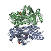
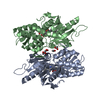
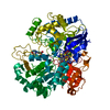
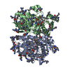
- PDB-6kr5: Crystal structure of O-Acetyl Serine Sulfhydrylase isoform 3 from... -
+ Open data

- Basic information
| Entry | Database: PDB / ID: 6kr5 | ||||||
|---|---|---|---|---|---|---|---|
| Title | Crystal structure of O-Acetyl Serine Sulfhydrylase isoform 3 from Entamoeba histolytica | ||||||
 Components Components | Cysteine synthase 3 | ||||||
 Keywords Keywords | TRANSFERASE / O-acetylserine sulphydralase / Cysteine synthase transferase O-acetyl-L-serine(thiol)lyase | ||||||
| Function / homology |  Function and homology informationcysteine synthase / hydrogen sulfide biosynthetic process / cysteine synthase activity / cysteine biosynthetic process from serine / transsulfuration / pyridoxal phosphate binding / cytoplasm Function and homology informationcysteine synthase / hydrogen sulfide biosynthetic process / cysteine synthase activity / cysteine biosynthetic process from serine / transsulfuration / pyridoxal phosphate binding / cytoplasmSimilarity search - Function | ||||||
| Biological species |   Entamoeba histolytica (eukaryote) Entamoeba histolytica (eukaryote) | ||||||
| Method | X-RAY DIFFRACTION / SYNCHROTRON / MOLECULAR REPLACEMENT / Resolution: 1.544 Å | ||||||
 Authors Authors | Dharavath, S. / Gourinath, S. | ||||||
 Citation Citation | Journal: Eur.J.Med.Chem. / Year: 2020 Title: Crystal structure of O-Acetylserine sulfhydralase (OASS) isoform 3 from Entamoeba histolytica: Pharmacophore-based virtual screening and validation of novel inhibitors. Authors: Dharavath, S. / Vijayan, R. / Kumari, K. / Tomar, P. / Gourinath, S. | ||||||
| History |
|
- Structure visualization
| Structure viewer | Molecule: MolmilJmol/JSmol |
|---|
- Downloads & links
-Download
| PDBx/mmCIF format | 6kr5.cif.gz | 156.4 KB | Display | PDBx/mmCIF format |
|---|---|---|---|---|
| PDB format | pdb6kr5.ent.gz | 120.6 KB | Display | PDB format |
| PDBx/mmJSON format | 6kr5.json.gz | Tree view | PDBx/mmJSON format | |
| Others |  Other downloads Other downloads |
-Validation report
| Arichive directory | https://data.pdbj.org/pub/pdb/validation_reports/kr/6kr5ftp://data.pdbj.org/pub/pdb/validation_reports/kr/6kr5 | HTTPS FTP |
|---|
-Related structure data
| Related structure data | 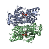 2pqmS S: Starting model for refinement |
|---|---|
| Similar structure data |


-Links
 PDBj
PDBj
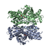
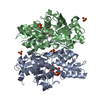
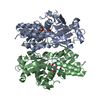
- Assembly
| Deposited unit | 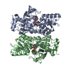
| ||||||||
|---|---|---|---|---|---|---|---|---|---|
| 1 |
| ||||||||
| Unit cell |
|
-Components
| #1: Protein | / Cysteine synthase A / putative / Cysteine synthase a putative Mass: 36491.160 Da / Num. of mol.: 2 Source method: isolated from a genetically manipulated source Source: (gene. exp.) Entamoeba histolytica (eukaryote) / Gene: CS 3, CL6EHI_060340, EHI_060340 / Production host:  Escherichia coli BL21(DE3) (bacteria) / Strain (production host): BL21(DE3) / References: UniProt: Q401L7, cysteine synthase Escherichia coli BL21(DE3) (bacteria) / Strain (production host): BL21(DE3) / References: UniProt: Q401L7, cysteine synthase#2: Chemical | Pyridoxal phosphate  Mass: 247.142 Da / Num. of mol.: 2 / Source method: obtained synthetically / Formula: C8H10NO6P / Feature type: SUBJECT OF INVESTIGATION Mass: 247.142 Da / Num. of mol.: 2 / Source method: obtained synthetically / Formula: C8H10NO6P / Feature type: SUBJECT OF INVESTIGATION#3: Water | ChemComp-HOH / | Water Mass: 18.015 Da / Num. of mol.: 704 / Source method: isolated from a natural source / Formula: H2O Mass: 18.015 Da / Num. of mol.: 704 / Source method: isolated from a natural source / Formula: H2OHas ligand of interest | Y | |
|---|
-Experimental details
-Experiment
| Experiment | Method: X-RAY DIFFRACTION / Number of used crystals: 1 |
|---|
- Sample preparation
| Crystal | Density Matthews: 2.18 Å3/Da / Density % sol: 43.46 % / Description: rod shaped |
|---|---|
| Crystal grow | Temperature: 289.15 K / Method: vapor diffusion / Details: 0.1M PCTP buffer, 25% PEG 1500 / PH range: 4.0 - 7.0 |
-Data collection
| Diffraction | Mean temperature: 193.15 K / Serial crystal experiment: N |
|---|---|
| Diffraction source | Source: SYNCHROTRON / Site: ESRF  / Beamline: ID30B / Wavelength: 1 Å / Beamline: ID30B / Wavelength: 1 Å |
| Detector | Type: MARMOSAIC 225 mm CCD / Detector: CCD / Date: Feb 6, 2016 |
| Radiation | Protocol: SINGLE WAVELENGTH / Monochromatic (M) / Laue (L): M / Scattering type: x-ray |
| Radiation wavelength | Wavelength: 1 Å / Relative weight: 1 |
| Reflection | Resolution: 1.54→50 Å / Num. obs: 92112 / % possible obs: 99.5 % / Redundancy: 7.3 % / CC1/2: 0.83 / Rpim(I) all: 0.27 / Rrim(I) all: 0.074 / Χ2: 2.1 / Net I/av σ(I): 2 / Net I/σ(I): 44 |
| Reflection shell | Resolution: 1.55→1.58 Å / Redundancy: 7 % / Mean I/σ(I) obs: 2 / Num. unique obs: 4544 / CC1/2: 0.83 / Rrim(I) all: 0.92 / Χ2: 0.7 / % possible all: 100 |
- Processing
| Software |
| ||||||||||||||||||||||||||||||||||||||||||||||||
|---|---|---|---|---|---|---|---|---|---|---|---|---|---|---|---|---|---|---|---|---|---|---|---|---|---|---|---|---|---|---|---|---|---|---|---|---|---|---|---|---|---|---|---|---|---|---|---|---|---|
| Refinement | Method to determine structure: MOLECULAR REPLACEMENT Starting model: 2PQM Resolution: 1.544→29.366 Å / SU ML: 0.14 / Cross valid method: THROUGHOUT / σ(F): 1.35 / Phase error: 23.82 / Stereochemistry target values: ML
| ||||||||||||||||||||||||||||||||||||||||||||||||
| Solvent computation | Shrinkage radii: 0.9 Å / VDW probe radii: 1.11 Å / Solvent model: FLAT BULK SOLVENT MODEL | ||||||||||||||||||||||||||||||||||||||||||||||||
| Displacement parameters | Biso max: 88.73 Å2 / Biso mean: 28.4354 Å2 / Biso min: 15.74 Å2 | ||||||||||||||||||||||||||||||||||||||||||||||||
| Refinement step | Cycle: final / Resolution: 1.544→29.366 Å
| ||||||||||||||||||||||||||||||||||||||||||||||||
| LS refinement shell | Refine-ID: X-RAY DIFFRACTION / Rfactor Rfree error: 0
|
