 Movie
Movie Controller

[English] 日本語
 Yorodumi
- PDB-4jbn: Crystal structure of O-Acetyl Serine Sulfhydrylase from Entamoeba... -
+ Open data

- Basic information
| Entry | Database: PDB / ID: 4jbn | ||||||
|---|---|---|---|---|---|---|---|
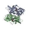
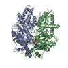
| Title | Crystal structure of O-Acetyl Serine Sulfhydrylase from Entamoeba histolytica in complex with Serine acetyl transferase derived tetrapeptide, SPSI | ||||||
 Components Components |
| ||||||
 Keywords Keywords | TRANSFERASE/TRANSFERASE INHIBITOR /  Cysteine Synthase / peptide inhibitor / PLP fold type 2 / Tryptophan synthase family / lyase / Serine Acetyl Transferase / TRANSFERASE-TRANSFERASE INHIBITOR complex Cysteine Synthase / peptide inhibitor / PLP fold type 2 / Tryptophan synthase family / lyase / Serine Acetyl Transferase / TRANSFERASE-TRANSFERASE INHIBITOR complex | ||||||
| Function / homology |  Function and homology information Function and homology information | ||||||
| Biological species |   Entamoeba histolytica (eukaryote) Entamoeba histolytica (eukaryote) | ||||||
| Method | X-RAY DIFFRACTION / MOLECULAR REPLACEMENT / Resolution: 1.87 Å | ||||||
 Authors Authors | Raj, I. / Gourinath, S. | ||||||
 Citation Citation | Journal: Biochim.Biophys.Acta / Year: 2013 Title: Molecular basis of ligand recognition by OASS from E. histolytica: insights from structural and molecular dynamics simulation studies Authors: Raj, I. / Mazumder, M. / Gourinath, S. | ||||||
| History |
|
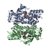
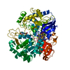
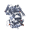
- Structure visualization
| Structure viewer | Molecule: MolmilJmol/JSmol |
|---|
- Downloads & links
-Download
| PDBx/mmCIF format | 4jbn.cif.gz | 146 KB | Display | PDBx/mmCIF format |
|---|---|---|---|---|
| PDB format | pdb4jbn.ent.gz | 114 KB | Display | PDB format |
| PDBx/mmJSON format | 4jbn.json.gz | Tree view | PDBx/mmJSON format | |
| Others |  Other downloads Other downloads |
-Validation report
| Arichive directory | https://data.pdbj.org/pub/pdb/validation_reports/jb/4jbnftp://data.pdbj.org/pub/pdb/validation_reports/jb/4jbn | HTTPS FTP |
|---|
-Related structure data
| Related structure data |  4il5C 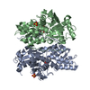 4jblC 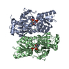 2pqmS S: Starting model for refinement C: citing same article ( |
|---|---|
| Similar structure data |


-Links
 PDBj
PDBj
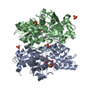
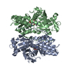
- Assembly
| Deposited unit | 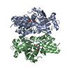
| ||||||||
|---|---|---|---|---|---|---|---|---|---|
| 1 |
| ||||||||
| Unit cell |
|
-Components
| #1: Protein | / O-Acetyl Serine Sulfhydrylase Mass: 37136.758 Da / Num. of mol.: 2 Source method: isolated from a genetically manipulated source Source: (gene. exp.) Entamoeba histolytica (eukaryote) / Strain: HM-1:IMSS / Gene: EhOASS / Plasmid: pET21c / Production host:  Escherichia coli (E. coli) / Strain (production host): BL21(DE3) / References: UniProt: O15570 Escherichia coli (E. coli) / Strain (production host): BL21(DE3) / References: UniProt: O15570#2: Protein/peptide | | Mass: 402.443 Da / Num. of mol.: 1 / Source method: obtained synthetically #3: Chemical | ChemComp-SO4 / | Sulfate  Mass: 96.063 Da / Num. of mol.: 1 / Source method: obtained synthetically / Formula: SO4 Mass: 96.063 Da / Num. of mol.: 1 / Source method: obtained synthetically / Formula: SO4#4: Water | ChemComp-HOH / | Water Mass: 18.015 Da / Num. of mol.: 251 / Source method: isolated from a natural source / Formula: H2O Mass: 18.015 Da / Num. of mol.: 251 / Source method: isolated from a natural source / Formula: H2O |
|---|
-Experimental details
-Experiment
| Experiment | Method: X-RAY DIFFRACTION / Number of used crystals: 1 |
|---|
- Sample preparation
| Crystal | Density Matthews: 2.44 Å3/Da / Density % sol: 49.68 % |
|---|---|
| Crystal grow | Temperature: 289 K / Method: vapor diffusion, hanging drop / pH: 7.2 Details: 2.1M AmSO4, 150mM NaCl, pH 7.2, VAPOR DIFFUSION, HANGING DROP, temperature 289K |
-Data collection
| Diffraction | Mean temperature: 100 K |
|---|---|
| Diffraction source | Source: ROTATING ANODE / Type: BRUKER AXS MICROSTAR / Wavelength: 1.54 Å |
| Detector | Type: MAR scanner 345 mm plate / Detector: IMAGE PLATE / Date: May 30, 2008 |
| Radiation | Monochromator: Cu / Protocol: SINGLE WAVELENGTH / Monochromatic (M) / Laue (L): M / Scattering type: x-ray |
| Radiation wavelength | Wavelength: 1.54 Å / Relative weight: 1 |
| Reflection | Resolution: 1.87→27.47 Å / Num. all: 58947 / Num. obs: 58645 / % possible obs: 94.6 % / Observed criterion σ(F): 5.3 / Observed criterion σ(I): 3.6 |
| Reflection shell | Resolution: 1.868→1.917 Å / % possible all: 90.77 |
- Processing
| Software |
| |||||||||||||||||||||||||||||||||||||||||||||
|---|---|---|---|---|---|---|---|---|---|---|---|---|---|---|---|---|---|---|---|---|---|---|---|---|---|---|---|---|---|---|---|---|---|---|---|---|---|---|---|---|---|---|---|---|---|---|
| Refinement | Method to determine structure: MOLECULAR REPLACEMENT Starting model: 2PQM Resolution: 1.87→27.47 Å / Cor.coef. Fo:Fc: 0.96 / Cor.coef. Fo:Fc free: 0.947 / Occupancy max: 1 / Occupancy min: 1 / SU B: 2.747 / SU ML: 0.085 / Cross valid method: THROUGHOUT / σ(F): 0 / ESU R: 0.14 / ESU R Free: 0.132 / Stereochemistry target values: MAXIMUM LIKELIHOOD Details: HYDROGENS HAVE BEEN USED IF PRESENT IN THE INPUT U VALUES: REFINED INDIVIDUALLY
| |||||||||||||||||||||||||||||||||||||||||||||
| Solvent computation | Ion probe radii: 0.8 Å / Shrinkage radii: 0.8 Å / VDW probe radii: 1.4 Å / Solvent model: MASK | |||||||||||||||||||||||||||||||||||||||||||||
| Displacement parameters | Biso max: 102.2 Å2 / Biso mean: 27.6853 Å2 / Biso min: 11.46 Å2
| |||||||||||||||||||||||||||||||||||||||||||||
| Refinement step | Cycle: LAST / Resolution: 1.87→27.47 Å
| |||||||||||||||||||||||||||||||||||||||||||||
| Refine LS restraints |
| |||||||||||||||||||||||||||||||||||||||||||||
| LS refinement shell | Resolution: 1.868→1.917 Å / Total num. of bins used: 20
|
