 Movie
Movie Controller

[English] 日本語
 Yorodumi
- PDB-6kqy: Crystal structure of human leucyl-tRNA synthetase, Leucine-bound form -
+ Open data

- Basic information
| Entry | Database: PDB / ID: 6kqy | ||||||
|---|---|---|---|---|---|---|---|
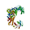
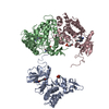
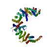
| Title | Crystal structure of human leucyl-tRNA synthetase, Leucine-bound form | ||||||
 Components Components | Leucine--tRNA ligase, cytoplasmic | ||||||
 Keywords Keywords |  LIGASE / leucyl-tRNA synthetase / leucine-bound structure LIGASE / leucyl-tRNA synthetase / leucine-bound structure | ||||||
| Function / homology |  Function and homology informationglutamine-tRNA ligase activity / glutaminyl-tRNA aminoacylation / Selenoamino acid metabolism / cellular response to leucine starvation / leucine-tRNA ligase / leucine-tRNA ligase activity / leucyl-tRNA aminoacylation / aminoacyl-tRNA synthetase multienzyme complex / Cytosolic tRNA aminoacylation / cellular response to L-leucine ...glutamine-tRNA ligase activity / glutaminyl-tRNA aminoacylation / Selenoamino acid metabolism / cellular response to leucine starvation / leucine-tRNA ligase / leucine-tRNA ligase activity / leucyl-tRNA aminoacylation / aminoacyl-tRNA synthetase multienzyme complex / Cytosolic tRNA aminoacylation / cellular response to L-leucine / tRNA aminoacylation for protein translation / aminoacyl-tRNA editing activity / regulation of cell size / positive regulation of TOR signaling / endomembrane system / positive regulation of TORC1 signaling / cellular response to amino acid starvation / GTPase activator activity / cellular response to amino acid stimulus / positive regulation of GTPase activity / lysosome / nuclear body / endoplasmic reticulum / ATP binding / cytosol / cytoplasm Function and homology informationglutamine-tRNA ligase activity / glutaminyl-tRNA aminoacylation / Selenoamino acid metabolism / cellular response to leucine starvation / leucine-tRNA ligase / leucine-tRNA ligase activity / leucyl-tRNA aminoacylation / aminoacyl-tRNA synthetase multienzyme complex / Cytosolic tRNA aminoacylation / cellular response to L-leucine ...glutamine-tRNA ligase activity / glutaminyl-tRNA aminoacylation / Selenoamino acid metabolism / cellular response to leucine starvation / leucine-tRNA ligase / leucine-tRNA ligase activity / leucyl-tRNA aminoacylation / aminoacyl-tRNA synthetase multienzyme complex / Cytosolic tRNA aminoacylation / cellular response to L-leucine / tRNA aminoacylation for protein translation / aminoacyl-tRNA editing activity / regulation of cell size / positive regulation of TOR signaling / endomembrane system / positive regulation of TORC1 signaling / cellular response to amino acid starvation / GTPase activator activity / cellular response to amino acid stimulus / positive regulation of GTPase activity / lysosome / nuclear body / endoplasmic reticulum / ATP binding / cytosol / cytoplasmSimilarity search - Function | ||||||
| Biological species |  Homo sapiens (human) Homo sapiens (human) | ||||||
| Method | X-RAY DIFFRACTION / SYNCHROTRON / MOLECULAR REPLACEMENT / Resolution: 3.3 Å | ||||||
 Authors Authors | Kim, S. / Son, J. / Kim, S. / Hwang, K.Y. | ||||||
 Citation Citation | Journal: Cell Rep / Year: 2021 Title: Leucine-sensing mechanism of leucyl-tRNA synthetase 1 for mTORC1 activation. Authors: Kim, S. / Yoon, I. / Son, J. / Park, J. / Kim, K. / Lee, J.H. / Park, S.Y. / Kang, B.S. / Han, J.M. / Hwang, K.Y. / Kim, S. | ||||||
| History |
|
- Structure visualization
| Structure viewer | Molecule: MolmilJmol/JSmol |
|---|
- Downloads & links
-Download
| PDBx/mmCIF format | 6kqy.cif.gz | 235.9 KB | Display | PDBx/mmCIF format |
|---|---|---|---|---|
| PDB format | pdb6kqy.ent.gz | 180.4 KB | Display | PDB format |
| PDBx/mmJSON format | 6kqy.json.gz | Tree view | PDBx/mmJSON format | |
| Others |  Other downloads Other downloads |
-Validation report
| Arichive directory | https://data.pdbj.org/pub/pdb/validation_reports/kq/6kqyftp://data.pdbj.org/pub/pdb/validation_reports/kq/6kqy | HTTPS FTP |
|---|
-Related structure data
| Related structure data |  6kidC  6kieC  6kr7C 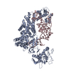 1wz2S  6ki4 S: Starting model for refinement C: citing same article ( |
|---|---|
| Similar structure data |


-Links
 PDBj
PDBj


- Assembly
| Deposited unit | 
| |||||||||
|---|---|---|---|---|---|---|---|---|---|---|
| 1 |
| |||||||||
| Unit cell |
| |||||||||
| Components on special symmetry positions |
|
-Components
| #1: Protein | Mass: 136055.359 Da / Num. of mol.: 1 Source method: isolated from a genetically manipulated source Details: SF file contains Friedel pairs / Source: (gene. exp.) Homo sapiens (human) / Gene: LARS, KIAA1352 / Production host:  Escherichia coli (E. coli) / References: UniProt: Q9P2J5, leucine-tRNA ligase Escherichia coli (E. coli) / References: UniProt: Q9P2J5, leucine-tRNA ligase | ||||||
|---|---|---|---|---|---|---|---|
| #2: Chemical | Leucine  Type: L-peptide linking / Mass: 131.173 Da / Num. of mol.: 2 / Source method: obtained synthetically / Formula: C6H13NO2 / Feature type: SUBJECT OF INVESTIGATION Type: L-peptide linking / Mass: 131.173 Da / Num. of mol.: 2 / Source method: obtained synthetically / Formula: C6H13NO2 / Feature type: SUBJECT OF INVESTIGATION#3: Chemical | ChemComp-PO4 / | Phosphate  Mass: 94.971 Da / Num. of mol.: 1 / Source method: obtained synthetically / Formula: PO4 Mass: 94.971 Da / Num. of mol.: 1 / Source method: obtained synthetically / Formula: PO4#4: Water | ChemComp-HOH / | Water Mass: 18.015 Da / Num. of mol.: 447 / Source method: isolated from a natural source / Formula: H2O Mass: 18.015 Da / Num. of mol.: 447 / Source method: isolated from a natural source / Formula: H2OHas ligand of interest | Y | |
-Experimental details
-Experiment
| Experiment | Method: X-RAY DIFFRACTION / Number of used crystals: 1 |
|---|
- Sample preparation
| Crystal | Density Matthews: 4.36 Å3/Da / Density % sol: 71.77 % |
|---|---|
| Crystal grow | Temperature: 293 K / Method: vapor diffusion, hanging drop Details: 0.1 M HEPES (pH 7.1), 0.42 M Ammonium sulfate, 24 % PEG3350 |
-Data collection
| Diffraction | Mean temperature: 273 K / Serial crystal experiment: N | |||||||||||||||||||||||||||||||||||||||||||||||||||||||||||||||||||||||||||||||||||||||||||||||||||||||||||||||||||||||||||||||||||||||||||||||||||||||||||||||||||||||||||||||||||||||||||||
|---|---|---|---|---|---|---|---|---|---|---|---|---|---|---|---|---|---|---|---|---|---|---|---|---|---|---|---|---|---|---|---|---|---|---|---|---|---|---|---|---|---|---|---|---|---|---|---|---|---|---|---|---|---|---|---|---|---|---|---|---|---|---|---|---|---|---|---|---|---|---|---|---|---|---|---|---|---|---|---|---|---|---|---|---|---|---|---|---|---|---|---|---|---|---|---|---|---|---|---|---|---|---|---|---|---|---|---|---|---|---|---|---|---|---|---|---|---|---|---|---|---|---|---|---|---|---|---|---|---|---|---|---|---|---|---|---|---|---|---|---|---|---|---|---|---|---|---|---|---|---|---|---|---|---|---|---|---|---|---|---|---|---|---|---|---|---|---|---|---|---|---|---|---|---|---|---|---|---|---|---|---|---|---|---|---|---|---|---|---|---|
| Diffraction source | Source: SYNCHROTRON / Site: PAL/PLS  / Beamline: 5C (4A) / Wavelength: 1 Å / Beamline: 5C (4A) / Wavelength: 1 Å | |||||||||||||||||||||||||||||||||||||||||||||||||||||||||||||||||||||||||||||||||||||||||||||||||||||||||||||||||||||||||||||||||||||||||||||||||||||||||||||||||||||||||||||||||||||||||||||
| Detector | Type: MAR CCD 165 mm / Detector: CCD / Date: Mar 25, 2018 | |||||||||||||||||||||||||||||||||||||||||||||||||||||||||||||||||||||||||||||||||||||||||||||||||||||||||||||||||||||||||||||||||||||||||||||||||||||||||||||||||||||||||||||||||||||||||||||
| Radiation | Protocol: SINGLE WAVELENGTH / Monochromatic (M) / Laue (L): M / Scattering type: x-ray | |||||||||||||||||||||||||||||||||||||||||||||||||||||||||||||||||||||||||||||||||||||||||||||||||||||||||||||||||||||||||||||||||||||||||||||||||||||||||||||||||||||||||||||||||||||||||||||
| Radiation wavelength | Wavelength: 1 Å / Relative weight: 1 | |||||||||||||||||||||||||||||||||||||||||||||||||||||||||||||||||||||||||||||||||||||||||||||||||||||||||||||||||||||||||||||||||||||||||||||||||||||||||||||||||||||||||||||||||||||||||||||
| Reflection | Resolution: 2.8→50 Å / Num. obs: 67806 / % possible obs: 93.5 % / Redundancy: 9.1 % / Rmerge(I) obs: 0.189 / Rpim(I) all: 0.06 / Rrim(I) all: 0.2 / Χ2: 2.155 / Net I/σ(I): 6.2 | |||||||||||||||||||||||||||||||||||||||||||||||||||||||||||||||||||||||||||||||||||||||||||||||||||||||||||||||||||||||||||||||||||||||||||||||||||||||||||||||||||||||||||||||||||||||||||||
| Reflection shell | Diffraction-ID: 1
|
- Processing
| Software |
| ||||||||||||||||||||||||||||||||||||||||||||||||||||||||||||||||||||||||||||||||||||||||||||||||||||||||||||||||||||||||||||||
|---|---|---|---|---|---|---|---|---|---|---|---|---|---|---|---|---|---|---|---|---|---|---|---|---|---|---|---|---|---|---|---|---|---|---|---|---|---|---|---|---|---|---|---|---|---|---|---|---|---|---|---|---|---|---|---|---|---|---|---|---|---|---|---|---|---|---|---|---|---|---|---|---|---|---|---|---|---|---|---|---|---|---|---|---|---|---|---|---|---|---|---|---|---|---|---|---|---|---|---|---|---|---|---|---|---|---|---|---|---|---|---|---|---|---|---|---|---|---|---|---|---|---|---|---|---|---|---|
| Refinement | Method to determine structure: MOLECULAR REPLACEMENT Starting model: 1wz2 Resolution: 3.3→49.76 Å / SU ML: 0.48 / Cross valid method: THROUGHOUT / σ(F): 1.5 / Phase error: 22.96 / Stereochemistry target values: ML
| ||||||||||||||||||||||||||||||||||||||||||||||||||||||||||||||||||||||||||||||||||||||||||||||||||||||||||||||||||||||||||||||
| Solvent computation | Shrinkage radii: 0.9 Å / VDW probe radii: 1.11 Å / Solvent model: FLAT BULK SOLVENT MODEL | ||||||||||||||||||||||||||||||||||||||||||||||||||||||||||||||||||||||||||||||||||||||||||||||||||||||||||||||||||||||||||||||
| Displacement parameters | Biso max: 225.72 Å2 / Biso mean: 48.6675 Å2 / Biso min: 9 Å2 | ||||||||||||||||||||||||||||||||||||||||||||||||||||||||||||||||||||||||||||||||||||||||||||||||||||||||||||||||||||||||||||||
| Refinement step | Cycle: final / Resolution: 3.3→49.76 Å
| ||||||||||||||||||||||||||||||||||||||||||||||||||||||||||||||||||||||||||||||||||||||||||||||||||||||||||||||||||||||||||||||
| LS refinement shell | Refine-ID: X-RAY DIFFRACTION / Rfactor Rfree error: 0 / Total num. of bins used: 17
|
