 Movie
Movie Controller

[English] 日本語
 Yorodumi
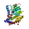
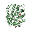
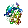
- PDB-6klr: Crystal structure of human WIPI3 in complex with the WIR-peptide ... -
+ Open data

- Basic information
| Entry | Database: PDB / ID: 6klr | ||||||
|---|---|---|---|---|---|---|---|
| Title | Crystal structure of human WIPI3 in complex with the WIR-peptide from ATG2A | ||||||
 Components Components | chimera ATG2A and WIPI3 | ||||||
 Keywords Keywords | LIPID BINDING PROTEIN / MEMBRANE BOUND PROTEIN | ||||||
| Function / homology |  Function and homology information Function and homology informationTSC1-TSC2 complex binding / organelle membrane contact site / phagophore /  protein lipidation / lipid transfer activity / glycophagy / nucleophagy / positive regulation of autophagosome assembly / autophagy of mitochondrion / protein localization to phagophore assembly site ...TSC1-TSC2 complex binding / organelle membrane contact site / phagophore / protein lipidation / lipid transfer activity / glycophagy / nucleophagy / positive regulation of autophagosome assembly / autophagy of mitochondrion / protein localization to phagophore assembly site / phagophore assembly site membrane / piecemeal microautophagy of the nucleus / phosphatidylinositol-3-phosphate binding / phagophore assembly site / reticulophagy / phosphatidylinositol-3,5-bisphosphate binding / Macroautophagy / extrinsic component of membrane / autophagosome assembly / protein-membrane adaptor activity / cellular response to starvation / lipid droplet / lysosome / endoplasmic reticulum membrane / cytosol protein lipidation / lipid transfer activity / glycophagy / nucleophagy / positive regulation of autophagosome assembly / autophagy of mitochondrion / protein localization to phagophore assembly site ...TSC1-TSC2 complex binding / organelle membrane contact site / phagophore / protein lipidation / lipid transfer activity / glycophagy / nucleophagy / positive regulation of autophagosome assembly / autophagy of mitochondrion / protein localization to phagophore assembly site / phagophore assembly site membrane / piecemeal microautophagy of the nucleus / phosphatidylinositol-3-phosphate binding / phagophore assembly site / reticulophagy / phosphatidylinositol-3,5-bisphosphate binding / Macroautophagy / extrinsic component of membrane / autophagosome assembly / protein-membrane adaptor activity / cellular response to starvation / lipid droplet / lysosome / endoplasmic reticulum membrane / cytosolSimilarity search - Function | ||||||
| Biological species |  Homo sapiens (human) Homo sapiens (human) | ||||||
| Method | X-RAY DIFFRACTION / SYNCHROTRON / MOLECULAR REPLACEMENT / Resolution: 2.21 Å | ||||||
 Authors Authors | Ren, J.Q. / Liang, R.B. / Feng, W. | ||||||
 Citation Citation | Journal: Nat Commun / Year: 2020 Title: Multi-site-mediated entwining of the linear WIR-motif around WIPI beta-propellers for autophagy. Authors: Ren, J. / Liang, R. / Wang, W. / Zhang, D. / Yu, L. / Feng, W. | ||||||
| History |
|
- Structure visualization
| Structure viewer | Molecule: MolmilJmol/JSmol |
|---|
- Downloads & links
-Download
| PDBx/mmCIF format | 6klr.cif.gz | 149 KB | Display | PDBx/mmCIF format |
|---|---|---|---|---|
| PDB format | pdb6klr.ent.gz | 112.7 KB | Display | PDB format |
| PDBx/mmJSON format | 6klr.json.gz | Tree view | PDBx/mmJSON format | |
| Others |  Other downloads Other downloads |
-Validation report
| Arichive directory | https://data.pdbj.org/pub/pdb/validation_reports/kl/6klrftp://data.pdbj.org/pub/pdb/validation_reports/kl/6klr | HTTPS FTP |
|---|
-Related structure data
| Related structure data |  6iyyS S: Starting model for refinement |
|---|---|
| Similar structure data |


-Links
 PDBj
PDBj- Assembly
| Deposited unit | 
| ||||||||
|---|---|---|---|---|---|---|---|---|---|
| 1 |
| ||||||||
| Unit cell |
|
-Components
| #1: Protein | Mass: 38459.828 Da / Num. of mol.: 2 / Mutation: Deletion of 75-80 and Deletion of 264-280 Source method: isolated from a genetically manipulated source Details: The sequence provided here is chimeric. We fused ATG2A (1374-1404) to the N-terminal of WIPI3. And we also deleted two loops in WIPI3 (Loop1: 75-80; loop2: 264-281). The first four residues ...Details: The sequence provided here is chimeric. We fused ATG2A (1374-1404) to the N-terminal of WIPI3. And we also deleted two loops in WIPI3 (Loop1: 75-80; loop2: 264-281). The first four residues (GPGS) is the expression tag. Residues form 5 to 35 (PRDGEPVVTQLHPGPIVVRDGYFSRPIGSTD) is the fused ATG2A sequence (from 1374 to 1404). Residues from 36 to 37 (GS) is the linker. The rest part is form WIPI3. Source: (gene. exp.) Homo sapiens (human) / Gene: ATG2A, KIAA0404, WDR45B, WDR45L, WIPI3 / Plasmid: pET32a / Production host:  Escherichia coli (E. coli) / References: UniProt: Q2TAZ0, UniProt: Q5MNZ6 Escherichia coli (E. coli) / References: UniProt: Q2TAZ0, UniProt: Q5MNZ6#2: Water | ChemComp-HOH / | Water Mass: 18.015 Da / Num. of mol.: 18 / Source method: isolated from a natural source / Formula: H2O Mass: 18.015 Da / Num. of mol.: 18 / Source method: isolated from a natural source / Formula: H2O |
|---|
-Experimental details
-Experiment
| Experiment | Method: X-RAY DIFFRACTION / Number of used crystals: 1 |
|---|
- Sample preparation
| Crystal | Density Matthews: 2.44 Å3/Da / Density % sol: 48.01 % Description: Authors state that Sfcheck gives the Matthews coefficient about 2.44, Phenix.Xtriage gives 2.18. |
|---|---|
| Crystal grow | Temperature: 289 K / Method: vapor diffusion, sitting drop / Details: 0.1M Tirs-HCl, pH 8.5, 20% (v/v) Ethanol / PH range: 8.5 |
-Data collection
| Diffraction | Mean temperature: 100 K / Serial crystal experiment: N |
|---|---|
| Diffraction source | Source: SYNCHROTRON / Site: SSRF  / Beamline: BL19U1 / Wavelength: 0.97855 Å / Beamline: BL19U1 / Wavelength: 0.97855 Å |
| Detector | Type: DECTRIS PILATUS3 6M / Detector: PIXEL / Date: Jan 18, 2018 |
| Radiation | Protocol: SINGLE WAVELENGTH / Monochromatic (M) / Laue (L): M / Scattering type: x-ray |
| Radiation wavelength | Wavelength: 0.97855 Å / Relative weight: 1 |
| Reflection | Resolution: 2.2→50 Å / Num. obs: 17842 / % possible obs: 99.3 % / Redundancy: 11.6 % / Biso Wilson estimate: 53.4 Å2 / CC1/2: 0.989 / Rmerge(I) obs: 0.093 / Rpim(I) all: 0.029 / Rrim(I) all: 0.102 / Χ2: 0.934 / Net I/av σ(I): 20.6 / Net I/σ(I): 23.1 |
| Reflection shell | Resolution: 2.2→2.28 Å / Redundancy: 7.9 % / Rmerge(I) obs: 0.423 / Mean I/σ(I) obs: 3.2 / Num. unique obs: 1650 / CC1/2: 0.925 / CC star: 0.98 / Rpim(I) all: 0.149 / Rrim(I) all: 0.451 / Χ2: 0.745 / % possible all: 96.2 |
- Processing
| Software |
| |||||||||||||||||||||||||||||||||||||||||||||||||||||||||||||||||||||||||||
|---|---|---|---|---|---|---|---|---|---|---|---|---|---|---|---|---|---|---|---|---|---|---|---|---|---|---|---|---|---|---|---|---|---|---|---|---|---|---|---|---|---|---|---|---|---|---|---|---|---|---|---|---|---|---|---|---|---|---|---|---|---|---|---|---|---|---|---|---|---|---|---|---|---|---|---|---|
| Refinement | Method to determine structure: MOLECULAR REPLACEMENT Starting model: 6IYY Resolution: 2.21→24.52 Å / SU ML: 0.36 / Cross valid method: THROUGHOUT / σ(F): 1.35 / Phase error: 36.27
| |||||||||||||||||||||||||||||||||||||||||||||||||||||||||||||||||||||||||||
| Solvent computation | Shrinkage radii: 0.9 Å / VDW probe radii: 1.11 Å | |||||||||||||||||||||||||||||||||||||||||||||||||||||||||||||||||||||||||||
| Displacement parameters | Biso max: 184.12 Å2 / Biso min: 30 Å2 | |||||||||||||||||||||||||||||||||||||||||||||||||||||||||||||||||||||||||||
| Refinement step | Cycle: final / Resolution: 2.21→24.52 Å
| |||||||||||||||||||||||||||||||||||||||||||||||||||||||||||||||||||||||||||
| LS refinement shell | Refine-ID: X-RAY DIFFRACTION / Rfactor Rfree error: 0 / Total num. of bins used: 6
| |||||||||||||||||||||||||||||||||||||||||||||||||||||||||||||||||||||||||||
| Refinement TLS params. | Method: refined / Refine-ID: X-RAY DIFFRACTION
| |||||||||||||||||||||||||||||||||||||||||||||||||||||||||||||||||||||||||||
| Refinement TLS group |
|
