 Movie
Movie Controller

[English] 日本語
 Yorodumi
- PDB-6kl0: Crystal structure of the S65T/F99S/M153T/V163A variant of perdeut... -
+ Open data

- Basic information
| Entry | Database: PDB / ID: 6kl0 | |||||||||
|---|---|---|---|---|---|---|---|---|---|---|
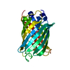
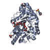
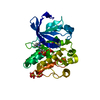
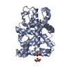
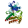
| Title | Crystal structure of the S65T/F99S/M153T/V163A variant of perdeuterated GFP at pD 7.0 | |||||||||
 Components Components | Green fluorescent protein | |||||||||
 Keywords Keywords | FLUORESCENT PROTEIN / Green Fluorescent Protein / visualization of hydrogen / perdeuteration / high resolution | |||||||||
| Function / homology | Green fluorescent protein, GFP / Green fluorescent protein-related / Green fluorescent protein / Green fluorescent protein / bioluminescence / generation of precursor metabolites and energy / DEUTERATED WATER / Green fluorescent protein Function and homology information Function and homology information | |||||||||
| Biological species |   Aequorea victoria (jellyfish) Aequorea victoria (jellyfish) | |||||||||
| Method | X-RAY DIFFRACTION / SYNCHROTRON / MOLECULAR REPLACEMENT / Resolution: 0.798 Å | |||||||||
 Authors Authors | Tai, Y. / Takaba, K. / Hanazono, Y. / Miki, K. / Takeda, K. | |||||||||
| Funding support |  Japan, 2items Japan, 2items
| |||||||||
 Citation Citation | Journal: Acta Crystallogr.,Sect.D / Year: 2019 Title: X-ray crystallographic studies on the hydrogen isotope effects of green fluorescent protein at sub-angstrom resolutions Authors: Tai, Y. / Takaba, K. / Hanazono, Y. / Dao, H.A. / Miki, K. / Takeda, K. | |||||||||
| History |
|
- Structure visualization
| Structure viewer | Molecule: MolmilJmol/JSmol |
|---|
- Downloads & links
-Download
| PDBx/mmCIF format | 6kl0.cif.gz | 213.1 KB | Display | PDBx/mmCIF format |
|---|---|---|---|---|
| PDB format | pdb6kl0.ent.gz | 174.3 KB | Display | PDB format |
| PDBx/mmJSON format | 6kl0.json.gz | Tree view | PDBx/mmJSON format | |
| Others |  Other downloads Other downloads |
-Validation report
| Arichive directory | https://data.pdbj.org/pub/pdb/validation_reports/kl/6kl0ftp://data.pdbj.org/pub/pdb/validation_reports/kl/6kl0 | HTTPS FTP |
|---|
-Related structure data
| Related structure data | 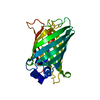 6kkzC 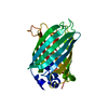 6kl1C 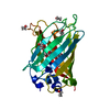 2wurS C: citing same article ( S: Starting model for refinement |
|---|---|
| Similar structure data |


-Links
 PDBj
PDBj


- Assembly
| Deposited unit | 
| ||||||||
|---|---|---|---|---|---|---|---|---|---|
| 1 |
| ||||||||
| Unit cell |
|
-Components
| #1: Protein | Mass: 25857.982 Da / Num. of mol.: 1 / Mutation: S65T, F99S, M153T, V163A Source method: isolated from a genetically manipulated source Source: (gene. exp.) Aequorea victoria (jellyfish) / Gene: GFP / Plasmid: pET21a / Production host:  Escherichia coli BL21(DE3) (bacteria) / References: UniProt: P42212 Escherichia coli BL21(DE3) (bacteria) / References: UniProt: P42212 |
|---|---|
| #2: Chemical | ChemComp-DOD / Heavy water  Mass: 18.015 Da / Num. of mol.: 606 / Source method: isolated from a natural source / Formula: D2O Mass: 18.015 Da / Num. of mol.: 606 / Source method: isolated from a natural source / Formula: D2O |
| Has ligand of interest | Y |
| Sequence details | (1) Residue SER 65 has been mutated to THR 65. Residues THR 65, TYR 66 and GLY 67 constitute the ...(1) Residue SER 65 has been mutated to THR 65. Residues THR 65, TYR 66 and GLY 67 constitute the chromophore CRO 66. The authors state that there are some difference in the structures between CRO in database and CRO in this model. They are in the terminal carboxylic and amino groups and the protonation of phenolic oxygen. (2) Q80R was caused by a PCR error in the early study (Chalfie, M. et al., Science, 1994). |
-Experimental details
-Experiment
| Experiment | Method: X-RAY DIFFRACTION / Number of used crystals: 1 |
|---|
- Sample preparation
| Crystal | Density Matthews: 2.11 Å3/Da / Density % sol: 41.76 % |
|---|---|
| Crystal grow | Temperature: 308 K / Method: vapor diffusion / pH: 6.6 / Details: PEG 4000, MgCl2, Tris-DCl buffer |
-Data collection
| Diffraction | Mean temperature: 50 K / Serial crystal experiment: Y |
|---|---|
| Diffraction source | Source: SYNCHROTRON / Site: SPring-8 / Beamline: BL44XU / Wavelength: 0.75 Å |
| Detector | Type: RAYONIX MX300HE / Detector: CCD / Date: Jul 21, 2017 |
| Radiation | Protocol: SINGLE WAVELENGTH / Monochromatic (M) / Laue (L): M / Scattering type: x-ray |
| Radiation wavelength | Wavelength: 0.75 Å / Relative weight: 1 |
| Reflection | Resolution: 0.798→50 Å / Num. obs: 229145 / % possible obs: 100 % / Redundancy: 6.7 % / Biso Wilson estimate: 6.16 Å2 / Rmerge(I) obs: 0.092 / Net I/σ(I): 29.6 |
| Reflection shell | Resolution: 0.798→0.81 Å / Redundancy: 6 % / Rmerge(I) obs: 1.459 / Mean I/σ(I) obs: 1.2 / Num. unique obs: 11357 / CC1/2: 0.475 / % possible all: 99.8 |
| Serial crystallography sample delivery | Method: fixed target |
- Processing
| Software |
| ||||||||||||||||||||||||||||||||||||||||||||||||||||||||||||||||||||||||||||||||||||||||||||||||||||||||||||||||||||||||||||||||||||||||||||||||||||||||||||||||||||||||||||||||||||||||||
|---|---|---|---|---|---|---|---|---|---|---|---|---|---|---|---|---|---|---|---|---|---|---|---|---|---|---|---|---|---|---|---|---|---|---|---|---|---|---|---|---|---|---|---|---|---|---|---|---|---|---|---|---|---|---|---|---|---|---|---|---|---|---|---|---|---|---|---|---|---|---|---|---|---|---|---|---|---|---|---|---|---|---|---|---|---|---|---|---|---|---|---|---|---|---|---|---|---|---|---|---|---|---|---|---|---|---|---|---|---|---|---|---|---|---|---|---|---|---|---|---|---|---|---|---|---|---|---|---|---|---|---|---|---|---|---|---|---|---|---|---|---|---|---|---|---|---|---|---|---|---|---|---|---|---|---|---|---|---|---|---|---|---|---|---|---|---|---|---|---|---|---|---|---|---|---|---|---|---|---|---|---|---|---|---|---|---|---|
| Refinement | Method to determine structure: MOLECULAR REPLACEMENT Starting model: 2WUR Resolution: 0.798→26.59 Å / SU ML: 0.08 / Cross valid method: THROUGHOUT / σ(F): 1.34 / Phase error: 13.13 / Stereochemistry target values: ML
| ||||||||||||||||||||||||||||||||||||||||||||||||||||||||||||||||||||||||||||||||||||||||||||||||||||||||||||||||||||||||||||||||||||||||||||||||||||||||||||||||||||||||||||||||||||||||||
| Solvent computation | Shrinkage radii: 0.9 Å / VDW probe radii: 1.11 Å / Solvent model: FLAT BULK SOLVENT MODEL | ||||||||||||||||||||||||||||||||||||||||||||||||||||||||||||||||||||||||||||||||||||||||||||||||||||||||||||||||||||||||||||||||||||||||||||||||||||||||||||||||||||||||||||||||||||||||||
| Displacement parameters | Biso max: 108.15 Å2 / Biso mean: 10.2835 Å2 / Biso min: 3.57 Å2 | ||||||||||||||||||||||||||||||||||||||||||||||||||||||||||||||||||||||||||||||||||||||||||||||||||||||||||||||||||||||||||||||||||||||||||||||||||||||||||||||||||||||||||||||||||||||||||
| Refinement step | Cycle: final / Resolution: 0.798→26.59 Å
| ||||||||||||||||||||||||||||||||||||||||||||||||||||||||||||||||||||||||||||||||||||||||||||||||||||||||||||||||||||||||||||||||||||||||||||||||||||||||||||||||||||||||||||||||||||||||||
| LS refinement shell | Refine-ID: X-RAY DIFFRACTION / Rfactor Rfree error: 0
|
