 Movie
Movie Controller

[English] 日本語
 Yorodumi
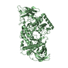
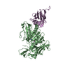
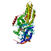
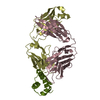
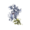
- PDB-6k7f: Crystal structure of MBPholo-Tim21 fusion protein with a 17-resid... -
+ Open data

- Basic information
| Entry | Database: PDB / ID: 6k7f | |||||||||
|---|---|---|---|---|---|---|---|---|---|---|
| Title | Crystal structure of MBPholo-Tim21 fusion protein with a 17-residue helical linker | |||||||||
 Components Components | Maltose/maltodextrin-binding periplasmic protein,Mitochondrial import inner membrane translocase subunit TIM21 | |||||||||
 Keywords Keywords | SUGAR BINDING PROTEIN /  TRANSLOCASE / MBP / Tim21 / fusion protein / helical linker TRANSLOCASE / MBP / Tim21 / fusion protein / helical linker | |||||||||
| Function / homology |  Function and homology information Function and homology informationTIM23 mitochondrial import inner membrane translocase complex / protein import into mitochondrial matrix / protein insertion into mitochondrial inner membrane / detection of maltose stimulus / maltose transport complex / maltose binding / maltose transport / maltodextrin transmembrane transport / carbohydrate transport / carbohydrate transmembrane transporter activity ...TIM23 mitochondrial import inner membrane translocase complex / protein import into mitochondrial matrix / protein insertion into mitochondrial inner membrane / detection of maltose stimulus / maltose transport complex / maltose binding / maltose transport / maltodextrin transmembrane transport / carbohydrate transport / carbohydrate transmembrane transporter activity / ATP-binding cassette (ABC) transporter complex, substrate-binding subunit-containing / ATP-binding cassette (ABC) transporter complex / cell chemotaxis / outer membrane-bounded periplasmic space / mitochondrial inner membrane / periplasmic space / DNA damage response / mitochondrion / membraneSimilarity search - Function | |||||||||
| Biological species |  Escherichia coli (E. coli) Escherichia coli (E. coli) Saccharomyces cerevisiae (brewer's yeast) Saccharomyces cerevisiae (brewer's yeast) | |||||||||
| Method | X-RAY DIFFRACTION / SYNCHROTRON / MOLECULAR REPLACEMENT / Resolution: 1.8 Å | |||||||||
 Authors Authors | Bala, S. / Shimada, A. / Kohda, D. | |||||||||
| Funding support |  Japan, 2items Japan, 2items
| |||||||||
 Citation Citation | Journal: Biochim Biophys Acta Gen Subj / Year: 2020 Title: Crystal contact-free conformation of an intrinsically flexible loop in protein crystal: Tim21 as the case study. Authors: Bala, S. / Shinya, S. / Srivastava, A. / Ishikawa, M. / Shimada, A. / Kobayashi, N. / Kojima, C. / Tama, F. / Miyashita, O. / Kohda, D. | |||||||||
| History |
|
- Structure visualization
| Structure viewer | Molecule: MolmilJmol/JSmol |
|---|
- Downloads & links
-Download
| PDBx/mmCIF format | 6k7f.cif.gz | 218.6 KB | Display | PDBx/mmCIF format |
|---|---|---|---|---|
| PDB format | pdb6k7f.ent.gz | 172.6 KB | Display | PDB format |
| PDBx/mmJSON format | 6k7f.json.gz | Tree view | PDBx/mmJSON format | |
| Others |  Other downloads Other downloads |
-Validation report
| Arichive directory | https://data.pdbj.org/pub/pdb/validation_reports/k7/6k7fftp://data.pdbj.org/pub/pdb/validation_reports/k7/6k7f | HTTPS FTP |
|---|
-Related structure data
| Related structure data |  6k7dC 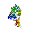 6k7eC 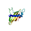 6k8qC  1anfS 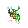 2ciuS S: Starting model for refinement C: citing same article ( |
|---|---|
| Similar structure data |


-Links
 PDBj
PDBj


- Assembly
| Deposited unit | 
| ||||||||
|---|---|---|---|---|---|---|---|---|---|
| 1 |
| ||||||||
| Unit cell |
|
-Components
| #1: Protein | Mass: 55648.258 Da / Num. of mol.: 1 / Mutation: A313V Source method: isolated from a genetically manipulated source Details: MBPholo-Tim21 fusion protein with a 17-residue helical linker Source: (gene. exp.) Escherichia coli (strain K12) (bacteria), (gene. exp.) Saccharomyces cerevisiae (strain ATCC 204508 / S288c) (yeast)Gene: malE, b4034, JW3994, TIM21, YGR033C / Production host: Escherichia coli BL21(DE3) (bacteria) / References: UniProt: P0AEX9, UniProt: P53220 |
|---|---|
| #2: Polysaccharide | alpha-D-glucopyranose-(1-4)-alpha-D-glucopyranose / alpha-maltose  , Oligosaccharide / Class: Nutrient / Mass: 342.297 Da / Num. of mol.: 1 , Oligosaccharide / Class: Nutrient / Mass: 342.297 Da / Num. of mol.: 1Source method: isolated from a genetically manipulated source Details: oligosaccharide / References: alpha-maltose |
| #3: Water | ChemComp-HOH / Water Mass: 18.015 Da / Num. of mol.: 319 / Source method: isolated from a natural source / Formula: H2O Mass: 18.015 Da / Num. of mol.: 319 / Source method: isolated from a natural source / Formula: H2O |
| Has ligand of interest | N |
-Experimental details
-Experiment
| Experiment | Method: X-RAY DIFFRACTION / Number of used crystals: 1 |
|---|
- Sample preparation
| Crystal | Density Matthews: 2.19 Å3/Da / Density % sol: 43.82 % |
|---|---|
| Crystal grow | Temperature: 293 K / Method: vapor diffusion, hanging drop / Details: 0.2M K/Na Tartrate, 16% PEG 3350 (w/v), microseeds |
-Data collection
| Diffraction | Mean temperature: 100 K / Serial crystal experiment: N |
|---|---|
| Diffraction source | Source: SYNCHROTRON / Site: SPring-8 / Beamline: BL44XU / Wavelength: 0.9 Å |
| Detector | Type: DECTRIS EIGER X 16M / Detector: PIXEL / Date: Nov 2, 2018 |
| Radiation | Protocol: SINGLE WAVELENGTH / Monochromatic (M) / Laue (L): M / Scattering type: x-ray |
| Radiation wavelength | Wavelength: 0.9 Å / Relative weight: 1 |
| Reflection | Resolution: 1.8→50 Å / Num. obs: 46285 / % possible obs: 99.9 % / Redundancy: 6.6 % / Biso Wilson estimate: 34.67 Å2 / Rmerge(I) obs: 0.047 / Net I/σ(I): 19.26 |
| Reflection shell | Resolution: 1.8→1.91 Å / Mean I/σ(I) obs: 2.02 / Num. unique obs: 7366 / CC1/2: 0.885 / % possible all: 99.8 |
- Processing
| Software |
| ||||||||||||||||||||||||||||||||||||||||||||||||||||||||||||||||||||||||||||||||||||||||||||||||||||||||||||||||||||||||||||||
|---|---|---|---|---|---|---|---|---|---|---|---|---|---|---|---|---|---|---|---|---|---|---|---|---|---|---|---|---|---|---|---|---|---|---|---|---|---|---|---|---|---|---|---|---|---|---|---|---|---|---|---|---|---|---|---|---|---|---|---|---|---|---|---|---|---|---|---|---|---|---|---|---|---|---|---|---|---|---|---|---|---|---|---|---|---|---|---|---|---|---|---|---|---|---|---|---|---|---|---|---|---|---|---|---|---|---|---|---|---|---|---|---|---|---|---|---|---|---|---|---|---|---|---|---|---|---|---|
| Refinement | Method to determine structure: MOLECULAR REPLACEMENT Starting model: 1ANF, 2CIU Resolution: 1.8→35.282 Å / SU ML: 0.2 / Cross valid method: FREE R-VALUE / σ(F): 1.37 / Phase error: 25.34 / Stereochemistry target values: ML
| ||||||||||||||||||||||||||||||||||||||||||||||||||||||||||||||||||||||||||||||||||||||||||||||||||||||||||||||||||||||||||||||
| Solvent computation | Shrinkage radii: 0.9 Å / VDW probe radii: 1.11 Å / Solvent model: FLAT BULK SOLVENT MODEL | ||||||||||||||||||||||||||||||||||||||||||||||||||||||||||||||||||||||||||||||||||||||||||||||||||||||||||||||||||||||||||||||
| Refinement step | Cycle: LAST / Resolution: 1.8→35.282 Å
| ||||||||||||||||||||||||||||||||||||||||||||||||||||||||||||||||||||||||||||||||||||||||||||||||||||||||||||||||||||||||||||||
| Refine LS restraints |
| ||||||||||||||||||||||||||||||||||||||||||||||||||||||||||||||||||||||||||||||||||||||||||||||||||||||||||||||||||||||||||||||
| LS refinement shell |
| ||||||||||||||||||||||||||||||||||||||||||||||||||||||||||||||||||||||||||||||||||||||||||||||||||||||||||||||||||||||||||||||
| Refinement TLS params. | Method: refined / Refine-ID: X-RAY DIFFRACTION
| ||||||||||||||||||||||||||||||||||||||||||||||||||||||||||||||||||||||||||||||||||||||||||||||||||||||||||||||||||||||||||||||
| Refinement TLS group |
|
