 Movie
Movie Controller

[English] 日本語
 Yorodumi
- PDB-2ciu: Structure of the IMS domain of the mitochondrial import protein T... -
+ Open data

- Basic information
| Entry | Database: PDB / ID: 2ciu | ||||||
|---|---|---|---|---|---|---|---|
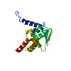
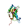
| Title | Structure of the IMS domain of the mitochondrial import protein Tim21 from S. cerevisiae | ||||||
 Components Components | IMPORT INNER MEMBRANE TRANSLOCASE SUBUNIT TIM21 MITOCHONDRIAL | ||||||
 Keywords Keywords |  PROTEIN TRANSPORT / MITOCHONDRIAL IMPORT / INNER MEMBRANE / MEMBRANE / MITOCHONDRION / TRANSIT PEPTIDE / TRANSLOCATION / TRANSMEMBRANE / TRANSPORT PROTEIN TRANSPORT / MITOCHONDRIAL IMPORT / INNER MEMBRANE / MEMBRANE / MITOCHONDRION / TRANSIT PEPTIDE / TRANSLOCATION / TRANSMEMBRANE / TRANSPORT | ||||||
| Function / homology |  Function and homology information Function and homology informationTIM23 mitochondrial import inner membrane translocase complex / protein import into mitochondrial matrix / protein insertion into mitochondrial inner membrane / mitochondrial inner membrane / mitochondrionSimilarity search - Function | ||||||
| Biological species |  SACCHAROMYCES CEREVISIAE (brewer's yeast) SACCHAROMYCES CEREVISIAE (brewer's yeast) | ||||||
| Method | X-RAY DIFFRACTION / SYNCHROTRON / MAD / Resolution: 1.6 Å | ||||||
 Authors Authors | Albrecht, R. / Zeth, K. / Rehling, P. / Pfanner, N. | ||||||
 Citation Citation | Journal: Embo Rep. / Year: 2006 Title: The Tim21 Binding Domain Connects the Preprotein Translocases of Both Mitochondrial Membranes Authors: Albrecht, R. / Rehling, P. / Chacinska, A. / Brix, J. / Cadamuro, S.A. / Volkmer, R. / Guiard, B. / Pfanner, N. / Zeth, K. | ||||||
| History |
| ||||||
| Remark 700 | SHEET THE SHEET STRUCTURE OF THIS MOLECULE IS BIFURCATED. IN ORDER TO REPRESENT THIS FEATURE IN ... SHEET THE SHEET STRUCTURE OF THIS MOLECULE IS BIFURCATED. IN ORDER TO REPRESENT THIS FEATURE IN THE SHEET RECORDS BELOW, TWO SHEETS ARE DEFINED. |
- Structure visualization
| Structure viewer | Molecule: MolmilJmol/JSmol |
|---|
- Downloads & links
-Download
| PDBx/mmCIF format | 2ciu.cif.gz | 41.1 KB | Display | PDBx/mmCIF format |
|---|---|---|---|---|
| PDB format | pdb2ciu.ent.gz | 28.4 KB | Display | PDB format |
| PDBx/mmJSON format | 2ciu.json.gz | Tree view | PDBx/mmJSON format | |
| Others |  Other downloads Other downloads |
-Validation report
| Arichive directory | https://data.pdbj.org/pub/pdb/validation_reports/ci/2ciuftp://data.pdbj.org/pub/pdb/validation_reports/ci/2ciu | HTTPS FTP |
|---|
-Related structure data
| Similar structure data |
|---|


-Links
 PDBj
PDBj- Assembly
| Deposited unit | 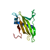
| ||||||||
|---|---|---|---|---|---|---|---|---|---|
| 1 |
| ||||||||
| Unit cell |
|
-Components
| #1: Protein | Mass: 14763.003 Da / Num. of mol.: 1 / Fragment: IMS DOMAIN, RESIDUES 103-225 Source method: isolated from a genetically manipulated source Source: (gene. exp.) SACCHAROMYCES CEREVISIAE (brewer's yeast)Organ: MITOCHONDRIUM / Plasmid: PPROEXHTA / Production host:  ESCHERICHIA COLI (E. coli) / Strain (production host): BL21(DE3) / References: UniProt: P53220 ESCHERICHIA COLI (E. coli) / Strain (production host): BL21(DE3) / References: UniProt: P53220 |
|---|---|
| #2: Water | ChemComp-HOH / Water Mass: 18.015 Da / Num. of mol.: 167 / Source method: isolated from a natural source / Formula: H2O Mass: 18.015 Da / Num. of mol.: 167 / Source method: isolated from a natural source / Formula: H2O |
| Compound details | ESSENTIAL COMPONENT OF THE TIM23 COMPLEX, A COMPLEX THAT MEDIATES THE TRANSLOCATION OF TRANSIT ...ESSENTIAL COMPONENT OF THE TIM23 COMPLEX, A COMPLEX THAT MEDIATES THE TRANSLOCAT |
-Experimental details
-Experiment
| Experiment | Method: X-RAY DIFFRACTION / Number of used crystals: 1 |
|---|
- Sample preparation
| Crystal | Density Matthews: 2.02 Å3/Da / Density % sol: 39 % |
|---|---|
| Crystal grow | pH: 6 / Details: 0.2 M LI2SO4, 0.1 M BIS-TRIS PH 6.5, 25% PEG 3350 |
-Data collection
| Diffraction | Mean temperature: 100 K |
|---|---|
| Diffraction source | Source: SYNCHROTRON / Site: MPG/DESY, HAMBURG  / Beamline: BW6 / Wavelength: 1.05 / Beamline: BW6 / Wavelength: 1.05 |
| Detector | Type: MARRESEARCH / Detector: CCD / Date: Jan 29, 2005 |
| Radiation | Protocol: SINGLE WAVELENGTH / Monochromatic (M) / Laue (L): M / Scattering type: x-ray |
| Radiation wavelength | Wavelength: 1.05 Å / Relative weight: 1 |
| Reflection | Resolution: 1.6→20 Å / Num. obs: 16515 / % possible obs: 97.5 % / Observed criterion σ(I): 3.72 / Redundancy: 3.33 % / Biso Wilson estimate: 21.4 Å2 / Rmerge(I) obs: 0.06 / Net I/σ(I): 18 |
| Reflection shell | Resolution: 1.58→1.68 Å / Redundancy: 3.27 % / Rmerge(I) obs: 0.33 / Mean I/σ(I) obs: 3.72 / % possible all: 94 |
- Processing
| Software |
| ||||||||||||||||||||||||||||||||||||||||||||||||||||||||||||
|---|---|---|---|---|---|---|---|---|---|---|---|---|---|---|---|---|---|---|---|---|---|---|---|---|---|---|---|---|---|---|---|---|---|---|---|---|---|---|---|---|---|---|---|---|---|---|---|---|---|---|---|---|---|---|---|---|---|---|---|---|---|
| Refinement | Method to determine structure: MAD / Resolution: 1.6→20 Å / Rfactor Rfree error: 0.009 / Data cutoff high absF: 909332.09 / Cross valid method: THROUGHOUT / σ(F): 0
| ||||||||||||||||||||||||||||||||||||||||||||||||||||||||||||
| Solvent computation | Bsol: 68.9141 Å2 / ksol: 0.419383 e/Å3 | ||||||||||||||||||||||||||||||||||||||||||||||||||||||||||||
| Displacement parameters | Biso mean: 22.9 Å2
| ||||||||||||||||||||||||||||||||||||||||||||||||||||||||||||
| Refine analyze |
| ||||||||||||||||||||||||||||||||||||||||||||||||||||||||||||
| Refinement step | Cycle: LAST / Resolution: 1.6→20 Å
| ||||||||||||||||||||||||||||||||||||||||||||||||||||||||||||
| Refine LS restraints |
| ||||||||||||||||||||||||||||||||||||||||||||||||||||||||||||
| LS refinement shell | Resolution: 1.6→1.7 Å / Rfactor Rfree error: 0.025 / Total num. of bins used: 6
| ||||||||||||||||||||||||||||||||||||||||||||||||||||||||||||
| Xplor file |
|
