[English] 日本語
Yorodumi
- PDB-6k6n: Crystal structure of SIVmac239 Nef protein -

+
Open data


ID or keywords:

Loading...

-
Basic information

Entry
Database: PDB / ID: 6k6n
TitleCrystal structure of SIVmac239 Nef protein
ComponentsProtein Nef
Keywords
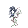
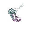
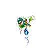
VIRAL PROTEIN / SIV / Nef / accesory protein
Function / homologyvirus-mediated perturbation of host defense response => GO:0019049 / HIV negative factor Nef / HIV-1 Nef protein, core domain superfamily / Negative factor, (F-Protein) or Nef / GTP binding / Protein Nef
Function and homology information
Biological speciesSimian immunodeficiency virus
MethodX-RAY DIFFRACTION / SYNCHROTRON / MOLECULAR REPLACEMENT / Resolution: 2.0002 Å
AuthorsHirao, K. / Andrews, S. / Kuroki, K. / Kusaka, H. / Tadokoro, T. / Kita, S. / Ose, T. / Rowland-Jones, S. / Maenaka, K.
Funding support Japan, 6items
OrganizationGrant numberCountry
Japan Society for the Promotion of Science15H02384 Japan
Japan Society for the Promotion of Science22121007 Japan
Japan Society for the Promotion of ScienceStrategic Young Researcher Overseas Visits Program for Accelerating Brain Circulation Japan
Japan Agency for Medical Research and Development (AMED)18am0101093j0002 Japan
Japan Agency for Medical Research and Development (AMED)18ae0101047h Japan
Japan Science and TechnologySupport Program for Implementation of New Equipment Sharing System Japan
CitationJournal: Iscience / Year: 2020
Title: Structure of HIV-2 Nef Reveals Features Distinct from HIV-1 Involved in Immune Regulation.
Authors: Hirao, K. / Andrews, S. / Kuroki, K. / Kusaka, H. / Tadokoro, T. / Kita, S. / Ose, T. / Rowland-Jones, S.L. / Maenaka, K.
History
DepositionJun 4, 2019Deposition site: PDBJ / Processing site: PDBJ
Revision 1.0Mar 25, 2020Provider: repository / Type: Initial release
Revision 1.1Apr 21, 2021Group: Database references / Category: citation / citation_author
Item: _citation.country / _citation.journal_abbrev ..._citation.country / _citation.journal_abbrev / _citation.journal_id_CSD / _citation.journal_id_ISSN / _citation.journal_volume / _citation.page_first / _citation.page_last / _citation.pdbx_database_id_DOI / _citation.pdbx_database_id_PubMed / _citation.title / _citation.year / _citation_author.name
Revision 1.2Nov 22, 2023Group: Data collection / Database references / Refinement description
Category: chem_comp_atom / chem_comp_bond ...chem_comp_atom / chem_comp_bond / database_2 / pdbx_initial_refinement_model
Item: _database_2.pdbx_DOI / _database_2.pdbx_database_accession

-
Structure visualization

Structure viewerMolecule:
MolmilJmol/JSmol

Downloads & links

-
Assembly

Deposited unit
A: Protein Nef


Theoretical massNumber of molelcules
Total (without water)20,5451
Polymers20,5451
Non-polymers00
Water1,72996
1


  • Idetical with deposited unit
  • defined by author
  • Evidence: gel filtration
TypeNameSymmetry operationNumber
identity operation1_555x,y,z1
Buried area0 Å2
ΔGint0 kcal/mol
Surface area9830 Å2
Unit cell
Length a, b, c (Å)72.531, 72.531, 111.087
Angle α, β, γ (deg.)90.000, 90.000, 120.000
Int Tables number179
Space group name H-MP6522
Space group name HallP652(x,y,z+1/12)

-
Components

#1: Protein Protein Nef


Mass: 20545.018 Da / Num. of mol.: 1
Source method: isolated from a genetically manipulated source
Source: (gene. exp.) Simian immunodeficiency virus / Gene: nef / Production host: Escherichia coli (E. coli) / References: UniProt: Q203Y4
#2: Water ChemComp-HOH / water / Water


Mass: 18.015 Da / Num. of mol.: 96 / Source method: isolated from a natural source / Formula: H2O

-
Experimental details

-
Experiment

ExperimentMethod: X-RAY DIFFRACTION / Number of used crystals: 1

-
Sample preparation

CrystalDensity Matthews: 2.02 Å3/Da / Density % sol: 39.09 %
Crystal growTemperature: 293 K / Method: vapor diffusion, sitting drop / pH: 5.6
Details: 0.1M Sodium citrate, 0.2M Ammonium acetate, 30% PEG4000

-
Data collection

DiffractionMean temperature: 100 K / Serial crystal experiment: N
Diffraction sourceSource: SYNCHROTRON / Site: Photon Factory / Beamline: BL-17A / Wavelength: 0.98 Å
DetectorType: DECTRIS PILATUS3 S 6M / Detector: PIXEL / Date: Jan 31, 2018
RadiationProtocol: SINGLE WAVELENGTH / Monochromatic (M) / Laue (L): M / Scattering type: x-ray
Radiation wavelengthWavelength: 0.98 Å / Relative weight: 1
ReflectionResolution: 2→41.61 Å / Num. obs: 12304 / % possible obs: 100 % / Redundancy: 18.7 % / Biso Wilson estimate: 29.1578178472 Å2 / CC1/2: 0.999 / Rmerge(I) obs: 0.112 / Rpim(I) all: 0.026 / Rrim(I) all: 0.115 / Net I/σ(I): 20 / Num. measured all: 229843 / Scaling rejects: 147
Reflection shell

Diffraction-ID: 1

Resolution (Å)Redundancy (%)Rmerge(I) obsNum. measured allNum. unique obsCC1/2Rpim(I) allRrim(I) allNet I/σ(I) obs% possible all
2-2.0518.91.035167058840.8740.2421.0633.2100
8.94-41.6113.30.019250418810.0050.01954.699.3

-
Processing

Software
NameVersionClassification
PHENIX1.11.1_2575refinement
Aimless0.5.32data scaling
PDB_EXTRACT3.24data extraction
XDSdata reduction
MOLREPphasing
RefinementMethod to determine structure: MOLECULAR REPLACEMENT
Starting model: 1AVV
Resolution: 2.0002→36.2655 Å / SU ML: 0.139412603568 / Cross valid method: THROUGHOUT / σ(F): 1.33785015254 / Phase error: 23.3477732977
Details: SF FILE CONTAINS FRIEDEL PAIRS UNDER I/F_MINUS AND I/F_PLUS COLUMNS.
RfactorNum. reflection% reflection
Rfree0.240531827051 596 4.86332109343 %
Rwork0.195196780635 --
obs0.197435856953 12255 99.9836827935 %
Solvent computationShrinkage radii: 0.9 Å / VDW probe radii: 1.11 Å
Displacement parametersBiso mean: 32.6391106576 Å2
Refinement stepCycle: LAST / Resolution: 2.0002→36.2655 Å
ProteinNucleic acidLigandSolventTotal
Num. atoms1263 0 0 96 1359
Refine LS restraints
Refine-IDTypeDev idealNumber
X-RAY DIFFRACTIONf_bond_d0.004001253765471299
X-RAY DIFFRACTIONf_angle_d0.5527280499051762
X-RAY DIFFRACTIONf_chiral_restr0.0454750802357180
X-RAY DIFFRACTIONf_plane_restr0.00376305158973225
X-RAY DIFFRACTIONf_dihedral_angle_d18.266097903484
LS refinement shell
Resolution (Å)Rfactor RfreeNum. reflection RfreeRfactor RworkNum. reflection RworkRefine-ID% reflection obs (%)
2.002-2.20150.2492231289811320.2006218457192852X-RAY DIFFRACTION100
2.2015-2.520.2815411609371500.1971564634252839X-RAY DIFFRACTION99.9665551839
2.52-3.17460.2397993711151580.2016878863752887X-RAY DIFFRACTION100
3.1746-36.26550.2277140519851560.1907426987383081X-RAY DIFFRACTION99.9691167387

+
About Yorodumi

-
News

-
Feb 9, 2022. New format data for meta-information of EMDB entries

New format data for meta-information of EMDB entries

  • Version 3 of the EMDB header file is now the official format.
  • The previous official version 1.9 will be removed from the archive.

Related info.:EMDB header

External links:wwPDB to switch to version 3 of the EMDB data model

-
Aug 12, 2020. Covid-19 info

Covid-19 info

URL: https://pdbj.org/emnavi/covid19.php

New page: Covid-19 featured information page in EM Navigator.

Related info.:Covid-19 info / Mar 5, 2020. Novel coronavirus structure data

+
Mar 5, 2020. Novel coronavirus structure data

Novel coronavirus structure data

Related info.:Yorodumi Speices / Aug 12, 2020. Covid-19 info

External links:COVID-19 featured content - PDBj / Molecule of the Month (242):Coronavirus Proteases

+
Jan 31, 2019. EMDB accession codes are about to change! (news from PDBe EMDB page)

EMDB accession codes are about to change! (news from PDBe EMDB page)

  • The allocation of 4 digits for EMDB accession codes will soon come to an end. Whilst these codes will remain in use, new EMDB accession codes will include an additional digit and will expand incrementally as the available range of codes is exhausted. The current 4-digit format prefixed with “EMD-” (i.e. EMD-XXXX) will advance to a 5-digit format (i.e. EMD-XXXXX), and so on. It is currently estimated that the 4-digit codes will be depleted around Spring 2019, at which point the 5-digit format will come into force.
  • The EM Navigator/Yorodumi systems omit the EMD- prefix.

Related info.:Q: What is EMD? / ID/Accession-code notation in Yorodumi/EM Navigator

External links:EMDB Accession Codes are Changing Soon! / Contact to PDBj

+
Jul 12, 2017. Major update of PDB

Major update of PDB

  • wwPDB released updated PDB data conforming to the new PDBx/mmCIF dictionary.
  • This is a major update changing the version number from 4 to 5, and with Remediation, in which all the entries are updated.
  • In this update, many items about electron microscopy experimental information are reorganized (e.g. em_software).
  • Now, EM Navigator and Yorodumi are based on the updated data.

External links:wwPDB Remediation / Enriched Model Files Conforming to OneDep Data Standards Now Available in the PDB FTP Archive

-
Yorodumi

Thousand views of thousand structures

  • Yorodumi is a browser for structure data from EMDB, PDB, SASBDB, etc.
  • This page is also the successor to EM Navigator detail page, and also detail information page/front-end page for Omokage search.
  • The word "yorodu" (or yorozu) is an old Japanese word meaning "ten thousand". "mi" (miru) is to see.

Related info.:EMDB / PDB / SASBDB / Comparison of 3 databanks / Yorodumi Search / Aug 31, 2016. New EM Navigator & Yorodumi / Yorodumi Papers / Jmol/JSmol / Function and homology information / Changes in new EM Navigator and Yorodumi

Read more