 Movie
Movie Controller

[English] 日本語
 Yorodumi
- PDB-6k4z: Single-chain Fv antibody of C6 COMPLEXED WITH 2-(4-HYDROXY-3-NITR... -
+ Open data

- Basic information
| Entry | Database: PDB / ID: 6k4z | ||||||
|---|---|---|---|---|---|---|---|
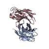
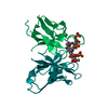
| Title | Single-chain Fv antibody of C6 COMPLEXED WITH 2-(4-HYDROXY-3-NITROPHENYL)ACETIC ACID | ||||||
 Components Components | scFv of C6 | ||||||
 Keywords Keywords |  IMMUNE SYSTEM / antigen binding / affinity maturation / Somatic hypermutation / conformational change IMMUNE SYSTEM / antigen binding / affinity maturation / Somatic hypermutation / conformational change | ||||||
| Function / homology | 2-(4-HYDROXY-3-NITROPHENYL)ACETIC ACID Function and homology information Function and homology information | ||||||
| Biological species |  Mus musculus (house mouse) Mus musculus (house mouse) | ||||||
| Method | X-RAY DIFFRACTION / SYNCHROTRON / MOLECULAR REPLACEMENT / Resolution: 1.65 Å | ||||||
 Authors Authors | Nishiguchi, A. / Numoto, N. / Ito, N. / Azuma, T. / Oda, M. | ||||||
 Citation Citation | Journal: Mol.Immunol. / Year: 2019 Title: Three-dimensional structure of a high affinity anti-(4-hydroxy-3-nitrophenyl)acetyl antibody possessing a glycine residue at position 95 of the heavy chain. Authors: Nishiguchi, A. / Numoto, N. / Ito, N. / Azuma, T. / Oda, M. | ||||||
| History |
|
- Structure visualization
| Structure viewer | Molecule: MolmilJmol/JSmol |
|---|
- Downloads & links
-Download
| PDBx/mmCIF format | 6k4z.cif.gz | 118.5 KB | Display | PDBx/mmCIF format |
|---|---|---|---|---|
| PDB format | pdb6k4z.ent.gz | 87 KB | Display | PDB format |
| PDBx/mmJSON format | 6k4z.json.gz | Tree view | PDBx/mmJSON format | |
| Others |  Other downloads Other downloads |
-Validation report
| Arichive directory | https://data.pdbj.org/pub/pdb/validation_reports/k4/6k4zftp://data.pdbj.org/pub/pdb/validation_reports/k4/6k4z | HTTPS FTP |
|---|
-Related structure data
| Related structure data |  3et9S S: Starting model for refinement |
|---|---|
| Similar structure data |


-Links
 PDBj
PDBj

- Assembly
| Deposited unit | 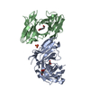
| ||||||||||||
|---|---|---|---|---|---|---|---|---|---|---|---|---|---|
| 1 | 
| ||||||||||||
| 2 | 
| ||||||||||||
| Unit cell |
|
-Components
| #1: Antibody | Mass: 26116.920 Da / Num. of mol.: 2 Source method: isolated from a genetically manipulated source Details: The fusion protein of expression tags (residues -2-0), the light chain (residues 1-108), linker GGGGSGGGGSGGGGS (residues 501-515), and the heavy chain (residues 1001-1113) Source: (gene. exp.) Mus musculus (house mouse) / Plasmid: pET-32TH / Production host:  Escherichia coli BL21(DE3) (bacteria) / Strain (production host): BL21(DE3) / Variant (production host): codon plus Escherichia coli BL21(DE3) (bacteria) / Strain (production host): BL21(DE3) / Variant (production host): codon plus#2: Chemical |   Mass: 197.145 Da / Num. of mol.: 2 / Source method: obtained synthetically / Formula: C8H7NO5 / Feature type: SUBJECT OF INVESTIGATION Mass: 197.145 Da / Num. of mol.: 2 / Source method: obtained synthetically / Formula: C8H7NO5 / Feature type: SUBJECT OF INVESTIGATION#3: Chemical | ChemComp-SO4 / | Sulfate  Mass: 96.063 Da / Num. of mol.: 1 / Source method: obtained synthetically / Formula: SO4 Mass: 96.063 Da / Num. of mol.: 1 / Source method: obtained synthetically / Formula: SO4#4: Chemical | ChemComp-GOL / | Glycerol  Mass: 92.094 Da / Num. of mol.: 1 / Source method: obtained synthetically / Formula: C3H8O3 Mass: 92.094 Da / Num. of mol.: 1 / Source method: obtained synthetically / Formula: C3H8O3#5: Water | ChemComp-HOH / | Water Mass: 18.015 Da / Num. of mol.: 542 / Source method: isolated from a natural source / Formula: H2O Mass: 18.015 Da / Num. of mol.: 542 / Source method: isolated from a natural source / Formula: H2O |
|---|
-Experimental details
-Experiment
| Experiment | Method: X-RAY DIFFRACTION / Number of used crystals: 1 |
|---|
- Sample preparation
| Crystal | Density Matthews: 2.37 Å3/Da / Density % sol: 48.13 % |
|---|---|
| Crystal grow | Temperature: 277 K / Method: vapor diffusion, hanging drop / pH: 8.5 Details: 0.14 M Lithium sulfate monohydrate, 0.07 M TRIS hydrochloride pH 8.5, 21% w/v Polyethylene glycol 4000 |
-Data collection
| Diffraction | Mean temperature: 100 K / Serial crystal experiment: N |
|---|---|
| Diffraction source | Source: SYNCHROTRON / Site: Photon Factory  / Beamline: BL-1A / Wavelength: 1.1 Å / Beamline: BL-1A / Wavelength: 1.1 Å |
| Detector | Type: DECTRIS EIGER X 4M / Detector: PIXEL / Date: Dec 12, 2018 |
| Radiation | Monochromator: Cryo-cooled channel-cut Si(111) / Protocol: SINGLE WAVELENGTH / Monochromatic (M) / Laue (L): M / Scattering type: x-ray |
| Radiation wavelength | Wavelength: 1.1 Å / Relative weight: 1 |
| Reflection | Resolution: 1.65→50 Å / Num. obs: 52628 / % possible obs: 96.2 % / Redundancy: 3.6 % / Biso Wilson estimate: 17.07 Å2 / CC1/2: 0.998 / Rsym value: 0.073 / Net I/σ(I): 11.2 |
| Reflection shell | Resolution: 1.65→1.75 Å / Redundancy: 3.5 % / Num. unique obs: 8339 / CC1/2: 0.716 / Rsym value: 0.586 / % possible all: 94.1 |
- Processing
| Software |
| ||||||||||||||||||||||||||||||||||||||||||||||||||||||||||||||||||||||||||||||||||||||||||||||||||||||||||||||||||||||||||||||||||||||||||||
|---|---|---|---|---|---|---|---|---|---|---|---|---|---|---|---|---|---|---|---|---|---|---|---|---|---|---|---|---|---|---|---|---|---|---|---|---|---|---|---|---|---|---|---|---|---|---|---|---|---|---|---|---|---|---|---|---|---|---|---|---|---|---|---|---|---|---|---|---|---|---|---|---|---|---|---|---|---|---|---|---|---|---|---|---|---|---|---|---|---|---|---|---|---|---|---|---|---|---|---|---|---|---|---|---|---|---|---|---|---|---|---|---|---|---|---|---|---|---|---|---|---|---|---|---|---|---|---|---|---|---|---|---|---|---|---|---|---|---|---|---|---|
| Refinement | Method to determine structure: MOLECULAR REPLACEMENT Starting model: 3ET9 Resolution: 1.65→43.29 Å / SU ML: 0.173 / Cross valid method: FREE R-VALUE / σ(F): 1.96 / Phase error: 21.1576
| ||||||||||||||||||||||||||||||||||||||||||||||||||||||||||||||||||||||||||||||||||||||||||||||||||||||||||||||||||||||||||||||||||||||||||||
| Solvent computation | Shrinkage radii: 0.9 Å / VDW probe radii: 1.11 Å | ||||||||||||||||||||||||||||||||||||||||||||||||||||||||||||||||||||||||||||||||||||||||||||||||||||||||||||||||||||||||||||||||||||||||||||
| Displacement parameters | Biso mean: 22.58 Å2 | ||||||||||||||||||||||||||||||||||||||||||||||||||||||||||||||||||||||||||||||||||||||||||||||||||||||||||||||||||||||||||||||||||||||||||||
| Refinement step | Cycle: LAST / Resolution: 1.65→43.29 Å
| ||||||||||||||||||||||||||||||||||||||||||||||||||||||||||||||||||||||||||||||||||||||||||||||||||||||||||||||||||||||||||||||||||||||||||||
| Refine LS restraints |
| ||||||||||||||||||||||||||||||||||||||||||||||||||||||||||||||||||||||||||||||||||||||||||||||||||||||||||||||||||||||||||||||||||||||||||||
| LS refinement shell |
|
