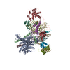
[English] 日本語
Yorodumi
- PDB-6k3f: Crystal Structure of beta-Arrestin 2 in Complex with CXCR7 Phosph... -

+
Open data


ID or keywords:

Loading...

-
Basic information

Entry
Database: PDB / ID: 6k3f
TitleCrystal Structure of beta-Arrestin 2 in Complex with CXCR7 Phosphopeptide
Components
  • Beta-arrestin-2Arrestin beta 2
  • Peptide from Atypical chemokine receptor 3
KeywordsSIGNALING PROTEIN / Complex
Function / homology
Function and homology information


type 2A serotonin receptor binding / platelet activating factor receptor binding / postsynaptic signal transduction / positive regulation of synaptic transmission, dopaminergic / oculomotor nerve development / alpha-1A adrenergic receptor binding / follicle-stimulating hormone receptor binding / Activation of SMO / G alpha (s) signalling events / alpha-1B adrenergic receptor binding ...type 2A serotonin receptor binding / platelet activating factor receptor binding / postsynaptic signal transduction / positive regulation of synaptic transmission, dopaminergic / oculomotor nerve development / alpha-1A adrenergic receptor binding / follicle-stimulating hormone receptor binding / Activation of SMO / G alpha (s) signalling events / alpha-1B adrenergic receptor binding / follicle-stimulating hormone signaling pathway / positive regulation of mesenchymal stem cell migration / angiotensin receptor binding / C-X-C chemokine binding / positive regulation of cardiac muscle cell differentiation / C-X-C chemokine receptor activity / WNT5A-dependent internalization of FZD4 / protein kinase B binding / MAP2K and MAPK activation / Ub-specific processing proteases / negative regulation of toll-like receptor signaling pathway / C-C chemokine receptor activity / Cargo recognition for clathrin-mediated endocytosis / C-C chemokine binding / Clathrin-mediated endocytosis / scavenger receptor activity / negative regulation of interleukin-12 production / regulation of G protein-coupled receptor signaling pathway / negative regulation of intrinsic apoptotic signaling pathway in response to DNA damage / positive regulation of calcium ion transport / arrestin family protein binding / G protein-coupled receptor internalization / type 1 angiotensin receptor binding / chemokine-mediated signaling pathway / adult walking behavior / Thrombin signalling through proteinase activated receptors (PARs) / Chemokine receptors bind chemokines / mitogen-activated protein kinase binding / positive regulation of epithelial cell apoptotic process / negative regulation of natural killer cell mediated cytotoxicity / negative regulation of interleukin-1 beta production / positive regulation of DNA biosynthetic process / negative regulation of release of cytochrome c from mitochondria / detection of temperature stimulus involved in sensory perception of pain / negative regulation of smooth muscle cell apoptotic process / negative regulation of interleukin-6 production / positive regulation of receptor internalization / endocytic vesicle / negative regulation of tumor necrosis factor production / D1 dopamine receptor binding / positive regulation of collagen biosynthetic process / vasculogenesis / coreceptor activity / negative regulation of canonical NF-kappaB signal transduction / clathrin-coated pit / negative regulation of protein ubiquitination / cell chemotaxis / transforming growth factor beta receptor signaling pathway / 14-3-3 protein binding / negative regulation of protein phosphorylation / G protein-coupled receptor binding / calcium-mediated signaling / regulation of protein phosphorylation / modulation of chemical synaptic transmission / receptor internalization / recycling endosome / endocytosis / positive regulation of peptidyl-tyrosine phosphorylation / protein transport / positive regulation of peptidyl-serine phosphorylation / positive regulation of cytosolic calcium ion concentration / G alpha (i) signalling events / cytoplasmic vesicle / postsynaptic membrane / proteasome-mediated ubiquitin-dependent protein catabolic process / basolateral plasma membrane / angiogenesis / negative regulation of neuron apoptotic process / dendritic spine / transcription by RNA polymerase II / positive regulation of ERK1 and ERK2 cascade / molecular adaptor activity / positive regulation of phosphatidylinositol 3-kinase/protein kinase B signal transduction / protein ubiquitination / early endosome / cell adhesion / endosome / immune response / positive regulation of protein phosphorylation / G protein-coupled receptor signaling pathway / protein domain specific binding / negative regulation of cell population proliferation / external side of plasma membrane / signaling receptor binding / intracellular membrane-bounded organelle / glutamatergic synapse / ubiquitin protein ligase binding / protein-containing complex binding / positive regulation of gene expression / enzyme binding
Similarity search - Function
Atypical chemokine receptor 3 / : / Arrestin, conserved site / Arrestins signature. / Arrestin / Arrestin, N-terminal / Arrestin-like, N-terminal / Arrestin C-terminal-like domain / Arrestin (or S-antigen), N-terminal domain / Arrestin (or S-antigen), C-terminal domain ...Atypical chemokine receptor 3 / : / Arrestin, conserved site / Arrestins signature. / Arrestin / Arrestin, N-terminal / Arrestin-like, N-terminal / Arrestin C-terminal-like domain / Arrestin (or S-antigen), N-terminal domain / Arrestin (or S-antigen), C-terminal domain / Arrestin (or S-antigen), C-terminal domain / Arrestin-like, C-terminal / G-protein coupled receptors family 1 signature. / G protein-coupled receptor, rhodopsin-like / GPCR, rhodopsin-like, 7TM / G-protein coupled receptors family 1 profile. / 7 transmembrane receptor (rhodopsin family) / Immunoglobulin E-set
Similarity search - Domain/homology
Atypical chemokine receptor 3 / Beta-arrestin-2
Similarity search - Component
Biological speciesRattus norvegicus (Norway rat)
Homo sapiens (human)
MethodX-RAY DIFFRACTION / SYNCHROTRON / MOLECULAR REPLACEMENT / Resolution: 2.3 Å
AuthorsMin, K.J. / Yoon, H.J. / Lee, H.H.
Funding support Korea, Republic Of, 3items
OrganizationGrant numberCountry
National Research Foundation (NRF, Korea)2015R1A5A1008958 Korea, Republic Of
National Research Foundation (NRF, Korea)2018R1A2B2008142 Korea, Republic Of
Ministry of Science, ICT and Future Planning (MSIP)2014M1A8A1049296 Korea, Republic Of
CitationJournal: Structure / Year: 2020
Title: Crystal Structure of beta-Arrestin 2 in Complex with CXCR7 Phosphopeptide.
Authors: Min, K. / Yoon, H.J. / Park, J.Y. / Baidya, M. / Dwivedi-Agnihotri, H. / Maharana, J. / Chaturvedi, M. / Chung, K.Y. / Shukla, A.K. / Lee, H.H.
History
DepositionMay 18, 2019Deposition site: PDBJ / Processing site: PDBJ
Revision 1.0Jun 10, 2020Provider: repository / Type: Initial release
Revision 1.1Jun 23, 2021Group: Database references / Structure summary / Category: citation / citation_author / struct
Item: _citation.country / _citation.journal_abbrev ..._citation.country / _citation.journal_abbrev / _citation.journal_id_ASTM / _citation.journal_id_CSD / _citation.journal_id_ISSN / _citation.journal_volume / _citation.page_first / _citation.page_last / _citation.pdbx_database_id_DOI / _citation.pdbx_database_id_PubMed / _citation.title / _citation.year / _struct.title
Revision 1.2Nov 22, 2023Group: Data collection / Database references / Refinement description
Category: chem_comp_atom / chem_comp_bond ...chem_comp_atom / chem_comp_bond / database_2 / pdbx_initial_refinement_model
Item: _database_2.pdbx_DOI / _database_2.pdbx_database_accession

-
Structure visualization

Structure viewerMolecule:
MolmilJmol/JSmol

Downloads & links

-
Assembly

Deposited unit
A: Beta-arrestin-2
U: Peptide from Atypical chemokine receptor 3
B: Beta-arrestin-2
V: Peptide from Atypical chemokine receptor 3
C: Beta-arrestin-2
W: Peptide from Atypical chemokine receptor 3
D: Beta-arrestin-2
X: Peptide from Atypical chemokine receptor 3
E: Beta-arrestin-2
Y: Peptide from Atypical chemokine receptor 3
F: Beta-arrestin-2
Z: Peptide from Atypical chemokine receptor 3


Theoretical massNumber of molelcules
Total (without water)266,30212
Polymers266,30212
Non-polymers00
Water9,440524
1


  • Idetical with deposited unit
  • defined by author&software
  • Evidence: light scattering
TypeNameSymmetry operationNumber
identity operation1_555x,y,z1
Buried area24640 Å2
ΔGint-127 kcal/mol
Surface area106150 Å2
MethodPISA
Unit cell
Length a, b, c (Å)91.170, 127.910, 206.040
Angle α, β, γ (deg.)90.000, 90.040, 90.000
Int Tables number5
Space group name H-MC121

-
Components

#1: Protein
Beta-arrestin-2 / Arrestin beta 2 / Arrestin beta-2


Mass: 42442.750 Da / Num. of mol.: 6
Source method: isolated from a genetically manipulated source
Source: (gene. exp.) Rattus norvegicus (Norway rat) / Gene: Arrb2 / Production host: Escherichia coli K-12 (bacteria) / References: UniProt: P29067
#2: Protein/peptide
Peptide from Atypical chemokine receptor 3 / / chemokine-related protein 1 / C-X-C chemokine receptor type 7 / CXCR-7 / Chemokine orphan receptor ...chemokine-related protein 1 / C-X-C chemokine receptor type 7 / CXCR-7 / Chemokine orphan receptor 1 / G-protein coupled receptor 159 / G-protein coupled receptor RDC1 homolog / RDC-1


Mass: 1940.975 Da / Num. of mol.: 6 / Source method: obtained synthetically / Source: (synth.) Homo sapiens (human) / References: UniProt: P25106
#3: Water ChemComp-HOH / water / Water


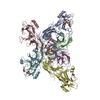
Mass: 18.015 Da / Num. of mol.: 524 / Source method: isolated from a natural source / Formula: H2O
Has ligand of interestY

-
Experimental details

-
Experiment

ExperimentMethod: X-RAY DIFFRACTION / Number of used crystals: 1

-
Sample preparation

CrystalDensity Matthews: 2.26 Å3/Da / Density % sol: 45.54 %
Crystal growTemperature: 295 K / Method: vapor diffusion, sitting drop
Details: 0.2M Ammonium acetate, 0.1M Bis-Tris pH5.5, 25% (w/v) PEG 3350

-
Data collection

DiffractionMean temperature: 100 K / Serial crystal experiment: N
Diffraction sourceSource: SYNCHROTRON / Site: SPring-8 / Beamline: BL26B1 / Wavelength: 1 Å
DetectorType: RAYONIX MX225HE / Detector: CCD / Date: Nov 3, 2018
RadiationProtocol: SINGLE WAVELENGTH / Monochromatic (M) / Laue (L): M / Scattering type: x-ray
Radiation wavelengthWavelength: 1 Å / Relative weight: 1
ReflectionResolution: 1.95→46.804 Å / Num. obs: 162425 / % possible obs: 99.67 % / Redundancy: 1 % / CC1/2: 0.711 / Net I/σ(I): 1.31
Reflection shellResolution: 1.95→2.07 Å / Num. unique obs: 49820 / CC1/2: 0.261

-
Processing

Software
NameVersionClassification
PHENIX1.13_2998refinement
XDSdata reduction
XDSdata scaling
MOLREPphasing
RefinementMethod to determine structure: MOLECULAR REPLACEMENT
Starting model: 5W0P
Resolution: 2.3→46.804 Å / Cross valid method: THROUGHOUT / σ(F): 0 / Phase error: 25.3 / Stereochemistry target values: TWIN_LSQ_F
RfactorNum. reflection% reflectionSelection details
Rfree0.2461 5310 5.07 %RANDOM
Rwork0.2332 99472 --
obs0.2358 104788 99.99 %-
Solvent computationShrinkage radii: 0.9 Å / VDW probe radii: 1.11 Å / Solvent model: FLAT BULK SOLVENT MODEL
Displacement parametersBiso max: 89.25 Å2 / Biso mean: 25.3474 Å2 / Biso min: 13.58 Å2
Refinement stepCycle: final / Resolution: 2.3→46.804 Å
ProteinNucleic acidLigandSolventTotal
Num. atoms16746 0 0 524 17270
Biso mean---19.75 -
Num. residues----2105
LS refinement shell

Refine-ID: X-RAY DIFFRACTION / Rfactor Rfree error: 0 / Total num. of bins used: 20

Resolution (Å)Rfactor RfreeNum. reflection RfreeRfactor RworkNum. reflection RworkNum. reflection all% reflection obs (%)
2.3001-2.33970.36232420.33454941518395
2.3397-2.38230.3182800.33564972525295
2.3823-2.42810.33172410.34284958519995
2.4281-2.47760.33322750.32024944521995
2.4776-2.53150.292650.31235043530895
2.5315-2.59040.33222590.30734896515595
2.5904-2.65520.27862540.3044946520095
2.6552-2.72690.30592560.28684986524295
2.7269-2.80720.27272600.26314980524095
2.8072-2.89780.25952730.25914959523295
2.8978-3.00130.27262490.24614948519795
3.0013-3.12150.25562410.23635012525395
3.1215-3.26350.24522820.22754970525295
3.2635-3.43550.22692680.22064943521195
3.4355-3.65060.23442980.20764957525594
3.6506-3.93240.22482690.19984980524995
3.9324-4.32780.2142720.17994974524695
4.3278-4.95340.18242990.17364943524294
4.9534-6.23820.22512400.21015044528495
6.2382-45.59390.25612860.22265076536295
Refinement TLS params.Method: refined / Origin x: 50.6348 Å / Origin y: 63.5994 Å / Origin z: 51.391 Å
111213212223313233
T0.1738 Å20.0024 Å20.0046 Å2-0.1899 Å2-0.0074 Å2--0.2867 Å2
L-0.015 °20.003 °2-0.0034 °2--0.0165 °2-0.0054 °2---0.0064 °2
S-0.008 Å °0.0056 Å °0.0072 Å °-0.0034 Å °0.0021 Å °-0.0099 Å °-0.0022 Å °-0.0004 Å °0.0042 Å °
Refinement TLS group
IDRefine-IDRefine TLS-IDSelection detailsAuth asym-IDAuth seq-ID
1X-RAY DIFFRACTION1allA8 - 350
2X-RAY DIFFRACTION1allU333 - 345
3X-RAY DIFFRACTION1allB8 - 346
4X-RAY DIFFRACTION1allV333 - 344
5X-RAY DIFFRACTION1allC8 - 350
6X-RAY DIFFRACTION1allW336 - 344
7X-RAY DIFFRACTION1allD8 - 350
8X-RAY DIFFRACTION1allX333 - 344
9X-RAY DIFFRACTION1allE8 - 346
10X-RAY DIFFRACTION1allY333 - 344
11X-RAY DIFFRACTION1allF8 - 350
12X-RAY DIFFRACTION1allZ336 - 344
13X-RAY DIFFRACTION1allS2 - 743

+
About Yorodumi

-
News

-
Feb 9, 2022. New format data for meta-information of EMDB entries

New format data for meta-information of EMDB entries

  • Version 3 of the EMDB header file is now the official format.
  • The previous official version 1.9 will be removed from the archive.

Related info.:EMDB header

External links:wwPDB to switch to version 3 of the EMDB data model

-
Aug 12, 2020. Covid-19 info

Covid-19 info

URL: https://pdbj.org/emnavi/covid19.php

New page: Covid-19 featured information page in EM Navigator.

Related info.:Covid-19 info / Mar 5, 2020. Novel coronavirus structure data

+
Mar 5, 2020. Novel coronavirus structure data

Novel coronavirus structure data

Related info.:Yorodumi Speices / Aug 12, 2020. Covid-19 info

External links:COVID-19 featured content - PDBj / Molecule of the Month (242):Coronavirus Proteases

+
Jan 31, 2019. EMDB accession codes are about to change! (news from PDBe EMDB page)

EMDB accession codes are about to change! (news from PDBe EMDB page)

  • The allocation of 4 digits for EMDB accession codes will soon come to an end. Whilst these codes will remain in use, new EMDB accession codes will include an additional digit and will expand incrementally as the available range of codes is exhausted. The current 4-digit format prefixed with “EMD-” (i.e. EMD-XXXX) will advance to a 5-digit format (i.e. EMD-XXXXX), and so on. It is currently estimated that the 4-digit codes will be depleted around Spring 2019, at which point the 5-digit format will come into force.
  • The EM Navigator/Yorodumi systems omit the EMD- prefix.

Related info.:Q: What is EMD? / ID/Accession-code notation in Yorodumi/EM Navigator

External links:EMDB Accession Codes are Changing Soon! / Contact to PDBj

+
Jul 12, 2017. Major update of PDB

Major update of PDB

  • wwPDB released updated PDB data conforming to the new PDBx/mmCIF dictionary.
  • This is a major update changing the version number from 4 to 5, and with Remediation, in which all the entries are updated.
  • In this update, many items about electron microscopy experimental information are reorganized (e.g. em_software).
  • Now, EM Navigator and Yorodumi are based on the updated data.

External links:wwPDB Remediation / Enriched Model Files Conforming to OneDep Data Standards Now Available in the PDB FTP Archive

-
Yorodumi

Thousand views of thousand structures

  • Yorodumi is a browser for structure data from EMDB, PDB, SASBDB, etc.
  • This page is also the successor to EM Navigator detail page, and also detail information page/front-end page for Omokage search.
  • The word "yorodu" (or yorozu) is an old Japanese word meaning "ten thousand". "mi" (miru) is to see.

Related info.:EMDB / PDB / SASBDB / Comparison of 3 databanks / Yorodumi Search / Aug 31, 2016. New EM Navigator & Yorodumi / Yorodumi Papers / Jmol/JSmol / Function and homology information / Changes in new EM Navigator and Yorodumi

Read more