 Movie
Movie Controller

[English] 日本語
 Yorodumi
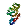
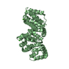
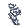
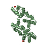
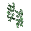
- PDB-6k22: Crystal structure of Ca-bound human Annexin A5 in low salt condition -
+ Open data

- Basic information
| Entry | Database: PDB / ID: 6k22 | ||||||
|---|---|---|---|---|---|---|---|
| Title | Crystal structure of Ca-bound human Annexin A5 in low salt condition | ||||||
 Components Components | Annexin A5 | ||||||
 Keywords Keywords | LIPID BINDING PROTEIN / Annexin A5 / phospholipid binding / membrane repair / AnxA5 | ||||||
| Function / homology |  Function and homology information Function and homology informationphospholipase inhibitor activity / endothelial microparticle / negative regulation of coagulation / calcium-dependent phospholipid binding / phosphatidylserine binding / : / phospholipid binding / sarcolemma / blood coagulation / Platelet degranulation ...phospholipase inhibitor activity / endothelial microparticle / negative regulation of coagulation / calcium-dependent phospholipid binding / phosphatidylserine binding / : / phospholipid binding / sarcolemma / blood coagulation / Platelet degranulation / collagen-containing extracellular matrix / external side of plasma membrane / focal adhesion / calcium ion binding / negative regulation of apoptotic process / signal transduction / extracellular exosome / extracellular region / membrane / cytosol / cytoplasmSimilarity search - Function | ||||||
| Biological species |  Homo sapiens (human) Homo sapiens (human) | ||||||
| Method | X-RAY DIFFRACTION / SYNCHROTRON / MOLECULAR REPLACEMENT / Resolution: 2.747 Å | ||||||
 Authors Authors | Hong, S. / Ha, N.-C. | ||||||
 Citation Citation | Journal: J.Struct.Biol. / Year: 2020 Title: High-resolution structures of annexin A5 in a two-dimensional array. Authors: Hong, S. / Na, S. / Kim, O.H. / Jeong, S. / Oh, B.C. / Ha, N.C. | ||||||
| History |
|
- Structure visualization
| Structure viewer | Molecule: MolmilJmol/JSmol |
|---|
- Downloads & links
-Download
| PDBx/mmCIF format | 6k22.cif.gz | 76.6 KB | Display | PDBx/mmCIF format |
|---|---|---|---|---|
| PDB format | pdb6k22.ent.gz | 55.1 KB | Display | PDB format |
| PDBx/mmJSON format | 6k22.json.gz | Tree view | PDBx/mmJSON format | |
| Others |  Other downloads Other downloads |
-Validation report
| Arichive directory | https://data.pdbj.org/pub/pdb/validation_reports/k2/6k22ftp://data.pdbj.org/pub/pdb/validation_reports/k2/6k22 | HTTPS FTP |
|---|
-Related structure data
| Related structure data | 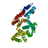 6k25C 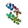 2xo3S C: citing same article ( S: Starting model for refinement |
|---|---|
| Similar structure data |


-Links
 PDBj
PDBj- Assembly
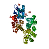
| Deposited unit | 
| ||||||||
|---|---|---|---|---|---|---|---|---|---|
| 1 |
| ||||||||
| Unit cell |
|
-Components
| #1: Protein | / Annexin-5 Mass: 37419.207 Da / Num. of mol.: 1 Source method: isolated from a genetically manipulated source Source: (gene. exp.) Homo sapiens (human) / Gene: ANXA5, ANX5 / Production host:  Escherichia coli BL21 (bacteria) / Strain (production host): BL21 / References: UniProt: P08758 Escherichia coli BL21 (bacteria) / Strain (production host): BL21 / References: UniProt: P08758 | ||
|---|---|---|---|
| #2: Chemical |   Mass: 40.078 Da / Num. of mol.: 3 / Source method: obtained synthetically / Formula: Ca Mass: 40.078 Da / Num. of mol.: 3 / Source method: obtained synthetically / Formula: Ca#3: Water | ChemComp-HOH / | Water Mass: 18.015 Da / Num. of mol.: 38 / Source method: isolated from a natural source / Formula: H2O Mass: 18.015 Da / Num. of mol.: 38 / Source method: isolated from a natural source / Formula: H2O |
-Experimental details
-Experiment
| Experiment | Method: X-RAY DIFFRACTION / Number of used crystals: 1 |
|---|
- Sample preparation
| Crystal | Density Matthews: 2.22 Å3/Da / Density % sol: 44.71 % |
|---|---|
| Crystal grow | Temperature: 287 K / Method: vapor diffusion, hanging drop Details: 0.2 M Potassium Citrate Tribasic pH 8.3 0.1 M HEPES pH 7.5 17% PEG3350 50uM CaCl2 2mM TCEP |
-Data collection
| Diffraction | Mean temperature: 193.15 K / Serial crystal experiment: N |
|---|---|
| Diffraction source | Source: SYNCHROTRON / Site: PAL/PLS  / Beamline: 5C (4A) / Wavelength: 1.2325 Å / Beamline: 5C (4A) / Wavelength: 1.2325 Å |
| Detector | Type: DECTRIS PILATUS 6M / Detector: PIXEL / Date: Apr 30, 2016 |
| Radiation | Protocol: SINGLE WAVELENGTH / Monochromatic (M) / Laue (L): M / Scattering type: x-ray |
| Radiation wavelength | Wavelength: 1.2325 Å / Relative weight: 1 |
| Reflection | Resolution: 2.747→50 Å / Num. obs: 9341 / % possible obs: 98.8 % / Redundancy: 16.8 % / Rmerge(I) obs: 0.145 / Rpim(I) all: 0.032 / Rrim(I) all: 0.149 / Χ2: 0.981 / Net I/σ(I): 14.8 |
| Reflection shell | Resolution: 2.75→2.8 Å / Redundancy: 7.4 % / Rmerge(I) obs: 0.39 / Mean I/σ(I) obs: 3.5 / Num. unique obs: 411 / CC1/2: 0.35 / Rpim(I) all: 0.134 / Rrim(I) all: 0.415 / % possible all: 87.4 |
- Processing
| Software |
| ||||||||||||||||||||||||||||
|---|---|---|---|---|---|---|---|---|---|---|---|---|---|---|---|---|---|---|---|---|---|---|---|---|---|---|---|---|---|
| Refinement | Method to determine structure: MOLECULAR REPLACEMENT Starting model: 2XO3 Resolution: 2.747→46.397 Å / SU ML: 0.41 / Cross valid method: FREE R-VALUE / σ(F): 1.58 / Phase error: 28.41
| ||||||||||||||||||||||||||||
| Solvent computation | Shrinkage radii: 0.9 Å / VDW probe radii: 1.11 Å | ||||||||||||||||||||||||||||
| Refinement step | Cycle: LAST / Resolution: 2.747→46.397 Å
| ||||||||||||||||||||||||||||
| Refine LS restraints |
| ||||||||||||||||||||||||||||
| LS refinement shell |
|
