 Movie
Movie Controller


+ Open data

- Basic information
| Entry | Database: PDB / ID: 2zoc | ||||||
|---|---|---|---|---|---|---|---|
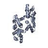
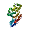
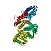
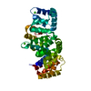
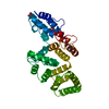
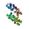
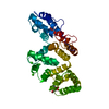
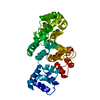
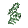
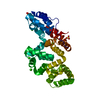
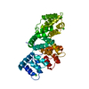
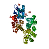
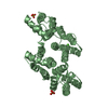
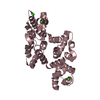
| Title | Crystal structure of recombinant human annexin IV | ||||||
 Components Components | Annexin A4 | ||||||
 Keywords Keywords | METAL BINDING PROTEIN / LIPID BINDING PROTEIN / helix / Annexin / Calcium / Calcium/phospholipid-binding | ||||||
| Function / homology |  Function and homology information Function and homology informationphospholipase inhibitor activity / negative regulation of interleukin-8 production / vesicle membrane / calcium-dependent phospholipid binding / negative regulation of NF-kappaB transcription factor activity / NF-kappaB binding / Notch signaling pathway / epithelial cell differentiation / calcium-dependent protein binding / nuclear membrane ...phospholipase inhibitor activity / negative regulation of interleukin-8 production / vesicle membrane / calcium-dependent phospholipid binding / negative regulation of NF-kappaB transcription factor activity / NF-kappaB binding / Notch signaling pathway / epithelial cell differentiation / calcium-dependent protein binding / nuclear membrane / collagen-containing extracellular matrix / calcium ion binding / regulation of transcription by RNA polymerase II / negative regulation of apoptotic process / perinuclear region of cytoplasm / cell surface / signal transduction / extracellular exosome / identical protein binding / nucleus / plasma membrane / cytoplasmSimilarity search - Function | ||||||
| Biological species |  Homo sapiens (human) Homo sapiens (human) | ||||||
| Method | X-RAY DIFFRACTION / SYNCHROTRON / MOLECULAR REPLACEMENT / Resolution: 2 Å | ||||||
 Authors Authors | Konno, M. / Kaneko-Kanzaki, Y. / Fushinobu-Okushi, N. / Mochizuki, K. / Uchikaw, E. / Satoh, A. / Aikawa, K. | ||||||
 Citation Citation | Journal: To be Published Title: The comparison of the loop structure of membrane binding sites between human and bovine annexin IV Authors: Konno, M. / Kaneko-Kanzaki, Y. / Fushinobu-Okushi, N. / Mochizuki, K. / Uchikawa, E. / Satoh, A. / Aikawa, K. | ||||||
| History |
|
- Structure visualization
| Structure viewer | Molecule: MolmilJmol/JSmol |
|---|
- Downloads & links
-Download
| PDBx/mmCIF format | 2zoc.cif.gz | 138.2 KB | Display | PDBx/mmCIF format |
|---|---|---|---|---|
| PDB format | pdb2zoc.ent.gz | 108.5 KB | Display | PDB format |
| PDBx/mmJSON format | 2zoc.json.gz | Tree view | PDBx/mmJSON format | |
| Others |  Other downloads Other downloads |
-Validation report
| Arichive directory | https://data.pdbj.org/pub/pdb/validation_reports/zo/2zocftp://data.pdbj.org/pub/pdb/validation_reports/zo/2zoc | HTTPS FTP |
|---|
-Related structure data
| Related structure data |  1annS S: Starting model for refinement |
|---|---|
| Similar structure data |


-Links
 PDBj
PDBj- Assembly
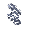
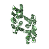
| Deposited unit | 
| ||||||||
|---|---|---|---|---|---|---|---|---|---|
| 1 | 
| ||||||||
| 2 | 
| ||||||||
| Unit cell |
|
-Components
| #1: Protein | / Annexin-4 / Annexin IV / Lipocortin IV / Endonexin I / Chromobindin-4 / Protein II / P32.5 / ...Annexin-4 / Annexin IV / Lipocortin IV / Endonexin I / Chromobindin-4 / Protein II / P32.5 / Placental anticoagulant protein II / PAP-II / PP4-X / 35-beta calcimedin / Carbohydrate-binding protein P33/P41 / P33/41 Mass: 35931.738 Da / Num. of mol.: 2 Source method: isolated from a genetically manipulated source Source: (gene. exp.) Homo sapiens (human) / Plasmid: pGEX-3X-annexinIV / Production host:  Escherichia coli (E. coli) / Strain (production host): HB101 / References: UniProt: P09525 Escherichia coli (E. coli) / Strain (production host): HB101 / References: UniProt: P09525#2: Chemical | ChemComp-CA /   Mass: 40.078 Da / Num. of mol.: 8 / Source method: obtained synthetically / Formula: Ca Mass: 40.078 Da / Num. of mol.: 8 / Source method: obtained synthetically / Formula: Ca#3: Water | ChemComp-HOH / | Water Mass: 18.015 Da / Num. of mol.: 224 / Source method: isolated from a natural source / Formula: H2O Mass: 18.015 Da / Num. of mol.: 224 / Source method: isolated from a natural source / Formula: H2O |
|---|
-Experimental details
-Experiment
| Experiment | Method: X-RAY DIFFRACTION / Number of used crystals: 1 |
|---|
- Sample preparation
| Crystal | Density Matthews: 2.16 Å3/Da / Density % sol: 42.95 % |
|---|---|
| Crystal grow | Temperature: 293 K / Method: vapor diffusion, hanging drop / pH: 7.5 Details: PEG 6000, calucium chloride, dioxisane, Tris-HCl, pH 7.5, VAPOR DIFFUSION, HANGING DROP, temperature 293K |
-Data collection
| Diffraction | Mean temperature: 100 K |
|---|---|
| Diffraction source | Source: SYNCHROTRON / Site: Photon Factory  / Beamline: BL-6A / Wavelength: 1 Å / Beamline: BL-6A / Wavelength: 1 Å |
| Detector | Type: ADSC QUANTUM 4 / Detector: CCD / Date: Jun 7, 1999 |
| Radiation | Monochromator: Si 111 CHANNEL / Protocol: SINGLE WAVELENGTH / Monochromatic (M) / Laue (L): M / Scattering type: x-ray |
| Radiation wavelength | Wavelength: 1 Å / Relative weight: 1 |
| Reflection | Resolution: 2→33.52 Å / Num. all: 38282 / Num. obs: 38282 / % possible obs: 92.1 % / Observed criterion σ(F): 0 / Observed criterion σ(I): 0 / Biso Wilson estimate: 17.3 Å2 / Rmerge(I) obs: 0.051 / Rsym value: 0.043 / Net I/σ(I): 13.2 |
| Reflection shell | Resolution: 2→2.11 Å / Rmerge(I) obs: 0.119 / Mean I/σ(I) obs: 6 / Num. unique all: 5405 / Rsym value: 0.119 / % possible all: 89.7 |
- Processing
| Software |
| |||||||||||||||||||||||||
|---|---|---|---|---|---|---|---|---|---|---|---|---|---|---|---|---|---|---|---|---|---|---|---|---|---|---|
| Refinement | Method to determine structure: MOLECULAR REPLACEMENT Starting model: PDB ENTRY 1ANN Resolution: 2→6 Å / Isotropic thermal model: Isotropic / Cross valid method: THROUGHOUT / σ(F): 1 / Stereochemistry target values: Engh & Huber
| |||||||||||||||||||||||||
| Refine analyze | Luzzati coordinate error obs: 0.25 Å / Luzzati d res low obs: 6 Å / Luzzati sigma a obs: 0.13 Å | |||||||||||||||||||||||||
| Refinement step | Cycle: LAST / Resolution: 2→6 Å
| |||||||||||||||||||||||||
| Refine LS restraints |
| |||||||||||||||||||||||||
| LS refinement shell | Resolution: 2→2.07 Å
|
