 ムービー
ムービー コントローラー


+ データを開く

- 基本情報
基本情報
| 登録情報 | データベース: PDB / ID: 6k08 | ||||||
|---|---|---|---|---|---|---|---|
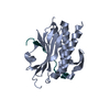
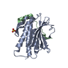
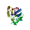
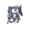
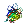
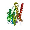
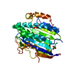
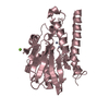
| タイトル | Crystal structure of REV7(R124A/A135D) in complex with a Shieldin3 fragment | ||||||
 要素 要素 |
| ||||||
 キーワード キーワード | TRANSCRIPTION/PROTEIN BINDING / DSB repair complex /  GENE REGULATION (遺伝子発現の調節) / TRANSCRIPTION-PROTEIN BINDING complex GENE REGULATION (遺伝子発現の調節) / TRANSCRIPTION-PROTEIN BINDING complex | ||||||
| 機能・相同性 |  機能・相同性情報 機能・相同性情報somatic diversification of immunoglobulins involved in immune response / DNA damage response, signal transduction resulting in transcription / telomere maintenance in response to DNA damage / negative regulation of transcription regulatory region DNA binding / zeta DNA polymerase complex / positive regulation of isotype switching / positive regulation of extracellular matrix assembly / negative regulation of transcription by competitive promoter binding / negative regulation of cell-cell adhesion mediated by cadherin / negative regulation of epithelial to mesenchymal transition ...somatic diversification of immunoglobulins involved in immune response / DNA damage response, signal transduction resulting in transcription / telomere maintenance in response to DNA damage / negative regulation of transcription regulatory region DNA binding / zeta DNA polymerase complex / positive regulation of isotype switching / positive regulation of extracellular matrix assembly / negative regulation of transcription by competitive promoter binding / negative regulation of cell-cell adhesion mediated by cadherin / negative regulation of epithelial to mesenchymal transition / JUN kinase binding / negative regulation of ubiquitin protein ligase activity / mitotic spindle assembly checkpoint signaling / positive regulation of double-strand break repair via nonhomologous end joining / negative regulation of double-strand break repair via homologous recombination / error-prone translesion synthesis / positive regulation of epithelial to mesenchymal transition / Translesion synthesis by REV1 / Translesion synthesis by POLK / Translesion synthesis by POLI / actin filament organization / regulation of cell growth / negative regulation of canonical Wnt signaling pathway / negative regulation of protein catabolic process / negative regulation of DNA-binding transcription factor activity / spindle / double-strand break repair / positive regulation of peptidyl-serine phosphorylation / site of double-strand break / 染色体 / RNA polymerase II-specific DNA-binding transcription factor binding / 細胞分裂 / DNA修復 / クロマチン / 核小体 / positive regulation of DNA-templated transcription / negative regulation of transcription by RNA polymerase II / 核質 / 細胞核 / 細胞質類似検索 - 分子機能 | ||||||
| 生物種 |  Homo sapiens (ヒト) Homo sapiens (ヒト) | ||||||
| 手法 | X線回折 / シンクロトロン / 分子置換 / 解像度: 2.312 Å | ||||||
 データ登録者 データ登録者 | Zhang, F. / Dai, Y. | ||||||
| 資金援助 |  中国, 1件 中国, 1件
| ||||||
 引用 引用 | ジャーナル: J.Biol.Chem. / 年: 2020 タイトル: Structural basis for shieldin complex subunit 3-mediated recruitment of the checkpoint protein REV7 during DNA double-strand break repair. 著者: Dai, Y. / Zhang, F. / Wang, L. / Shan, S. / Gong, Z. / Zhou, Z. | ||||||
| 履歴 |
|
- 構造の表示
構造の表示
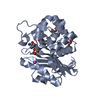
| 構造ビューア | 分子: MolmilJmol/JSmol |
|---|
- ダウンロードとリンク
-ダウンロード
| PDBx/mmCIF形式 | 6k08.cif.gz | 105.8 KB | 表示 | PDBx/mmCIF形式 |
|---|---|---|---|---|
| PDB形式 | pdb6k08.ent.gz | 79.5 KB | 表示 | PDB形式 |
| PDBx/mmJSON形式 | 6k08.json.gz | ツリー表示 | PDBx/mmJSON形式 | |
| その他 |  その他のダウンロード その他のダウンロード |
-検証レポート
| アーカイブディレクトリ | https://data.pdbj.org/pub/pdb/validation_reports/k0/6k08ftp://data.pdbj.org/pub/pdb/validation_reports/k0/6k08 | HTTPS FTP |
|---|
-関連構造データ


-リンク
 PDBj
PDBj


- 集合体
集合体
| 登録構造単位 | 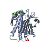
| ||||||||
|---|---|---|---|---|---|---|---|---|---|
| 1 |
| ||||||||
| 単位格子 |
|
-要素
| #1: タンパク質 | 分子量: 25239.197 Da / 分子数: 1 / 変異: R124A,A135D / 由来タイプ: 組換発現 / 由来: (組換発現) Homo sapiens (ヒト) / 遺伝子: MAD2L2, MAD2B, REV7 / 発現宿主:  Escherichia coli K-12 (大腸菌) / 株 (発現宿主): K-12 / 参照: UniProt: Q9UI95 Escherichia coli K-12 (大腸菌) / 株 (発現宿主): K-12 / 参照: UniProt: Q9UI95 |
|---|---|
| #2: タンパク質・ペプチド | 分子量: 3358.924 Da / 分子数: 1 / 由来タイプ: 組換発現 / 由来: (組換発現) Homo sapiens (ヒト) / 遺伝子: SHLD3, FLJ26957, RINN1 / 発現宿主: Escherichia coli K-12 (大腸菌) / 株 (発現宿主): K-12 / 参照: UniProt: Q6ZNX1 |
| #3: 化合物 | ChemComp-SO4 / 硫酸塩  分子量: 96.063 Da / 分子数: 1 / 由来タイプ: 合成 / 式: SO4 分子量: 96.063 Da / 分子数: 1 / 由来タイプ: 合成 / 式: SO4 |
| #4: 水 | ChemComp-HOH / 水 分子量: 18.015 Da / 分子数: 14 / 由来タイプ: 天然 / 式: H2O 分子量: 18.015 Da / 分子数: 14 / 由来タイプ: 天然 / 式: H2O |
-実験情報
-実験
| 実験 | 手法: X線回折 / 使用した結晶の数: 1 |
|---|
- 試料調製
試料調製
| 結晶 | マシュー密度: 2.49 Å3/Da / 溶媒含有率: 50.72 % |
|---|---|
| 結晶化 | 温度: 289 K / 手法: 蒸気拡散法, ハンギングドロップ法 / 詳細: sodium citrate tribasic, ammonium sulfate |
-データ収集
| 回折 | 平均測定温度: 80 K / Serial crystal experiment: N |
|---|---|
| 放射光源 | 由来: シンクロトロン / サイト: SSRF / ビームライン: BL19U1 / 波長: 0.987 Å |
| 検出器 | タイプ: DECTRIS PILATUS 6M-F / 検出器: PIXEL / 日付: 2018年12月3日 |
| 放射 | プロトコル: SINGLE WAVELENGTH / 単色(M)・ラウエ(L): M / 散乱光タイプ: x-ray |
| 放射波長 | 波長: 0.987 Å / 相対比: 1 |
| 反射 | 解像度: 2.31→44.49 Å / Num. obs: 13088 / % possible obs: 99.9 % / 冗長度: 9.3 % / Rpim(I) all: 0.025 / Net I/σ(I): 16.7 |
| 反射 シェル | 解像度: 2.31→2.39 Å / 冗長度: 9.8 % / Mean I/σ(I) obs: 2.4 / Num. unique obs: 1283 / Rpim(I) all: 0.453 / % possible all: 99.9 |
- 解析
解析
| ソフトウェア |
| |||||||||||||||||||||||||||||||||||||||||||||||||||||||||||||||||||||||||||
|---|---|---|---|---|---|---|---|---|---|---|---|---|---|---|---|---|---|---|---|---|---|---|---|---|---|---|---|---|---|---|---|---|---|---|---|---|---|---|---|---|---|---|---|---|---|---|---|---|---|---|---|---|---|---|---|---|---|---|---|---|---|---|---|---|---|---|---|---|---|---|---|---|---|---|---|---|
| 精密化 | 構造決定の手法: 分子置換 開始モデル: 3ABD 解像度: 2.312→44.493 Å / 交差検証法: FREE R-VALUE / σ(F): 1.35
| |||||||||||||||||||||||||||||||||||||||||||||||||||||||||||||||||||||||||||
| 溶媒の処理 | 減衰半径: 0.9 Å / VDWプローブ半径: 1.11 Å | |||||||||||||||||||||||||||||||||||||||||||||||||||||||||||||||||||||||||||
| 精密化ステップ | サイクル: LAST / 解像度: 2.312→44.493 Å
| |||||||||||||||||||||||||||||||||||||||||||||||||||||||||||||||||||||||||||
| 拘束条件 |
| |||||||||||||||||||||||||||||||||||||||||||||||||||||||||||||||||||||||||||
| LS精密化 シェル |
| |||||||||||||||||||||||||||||||||||||||||||||||||||||||||||||||||||||||||||
| 精密化 TLS | 手法: refined / Refine-ID: X-RAY DIFFRACTION
| |||||||||||||||||||||||||||||||||||||||||||||||||||||||||||||||||||||||||||
| 精密化 TLSグループ |
|
