 Movie
Movie Controller

[English] 日本語
 Yorodumi
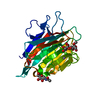
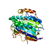
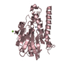
- PDB-6k08: Crystal structure of REV7(R124A/A135D) in complex with a Shieldin... -
+ Open data

- Basic information
| Entry | Database: PDB / ID: 6k08 | ||||||
|---|---|---|---|---|---|---|---|
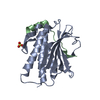
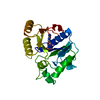
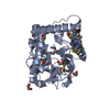
| Title | Crystal structure of REV7(R124A/A135D) in complex with a Shieldin3 fragment | ||||||
 Components Components |
| ||||||
 Keywords Keywords | TRANSCRIPTION/PROTEIN BINDING / DSB repair complex /  GENE REGULATION / TRANSCRIPTION-PROTEIN BINDING complex GENE REGULATION / TRANSCRIPTION-PROTEIN BINDING complex | ||||||
| Function / homology |  Function and homology information Function and homology informationsomatic diversification of immunoglobulins involved in immune response / DNA damage response, signal transduction resulting in transcription / telomere maintenance in response to DNA damage / negative regulation of transcription regulatory region DNA binding / zeta DNA polymerase complex / positive regulation of isotype switching / positive regulation of extracellular matrix assembly / negative regulation of epithelial to mesenchymal transition / negative regulation of transcription by competitive promoter binding / negative regulation of cell-cell adhesion mediated by cadherin ...somatic diversification of immunoglobulins involved in immune response / DNA damage response, signal transduction resulting in transcription / telomere maintenance in response to DNA damage / negative regulation of transcription regulatory region DNA binding / zeta DNA polymerase complex / positive regulation of isotype switching / positive regulation of extracellular matrix assembly / negative regulation of epithelial to mesenchymal transition / negative regulation of transcription by competitive promoter binding / negative regulation of cell-cell adhesion mediated by cadherin / JUN kinase binding / negative regulation of ubiquitin protein ligase activity / negative regulation of double-strand break repair via homologous recombination / mitotic spindle assembly checkpoint signaling / positive regulation of double-strand break repair via nonhomologous end joining / positive regulation of epithelial to mesenchymal transition / error-prone translesion synthesis / Translesion synthesis by REV1 / Translesion synthesis by POLK / Translesion synthesis by POLI / actin filament organization / regulation of cell growth / negative regulation of canonical Wnt signaling pathway / negative regulation of DNA-binding transcription factor activity / negative regulation of protein catabolic process / spindle / double-strand break repair / positive regulation of peptidyl-serine phosphorylation / site of double-strand break / chromosome / RNA polymerase II-specific DNA-binding transcription factor binding / cell division / DNA repair / chromatin / nucleolus / positive regulation of DNA-templated transcription / negative regulation of transcription by RNA polymerase II / nucleoplasm / nucleus / cytoplasmSimilarity search - Function | ||||||
| Biological species |  Homo sapiens (human) Homo sapiens (human) | ||||||
| Method | X-RAY DIFFRACTION / SYNCHROTRON / MOLECULAR REPLACEMENT / Resolution: 2.312 Å | ||||||
 Authors Authors | Zhang, F. / Dai, Y. | ||||||
| Funding support |  China, 1items China, 1items
| ||||||
 Citation Citation | Journal: J.Biol.Chem. / Year: 2020 Title: Structural basis for shieldin complex subunit 3-mediated recruitment of the checkpoint protein REV7 during DNA double-strand break repair. Authors: Dai, Y. / Zhang, F. / Wang, L. / Shan, S. / Gong, Z. / Zhou, Z. | ||||||
| History |
|
- Structure visualization
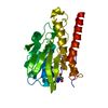
| Structure viewer | Molecule: MolmilJmol/JSmol |
|---|
- Downloads & links
-Download
| PDBx/mmCIF format | 6k08.cif.gz | 105.8 KB | Display | PDBx/mmCIF format |
|---|---|---|---|---|
| PDB format | pdb6k08.ent.gz | 79.5 KB | Display | PDB format |
| PDBx/mmJSON format | 6k08.json.gz | Tree view | PDBx/mmJSON format | |
| Others |  Other downloads Other downloads |
-Validation report
| Arichive directory | https://data.pdbj.org/pub/pdb/validation_reports/k0/6k08ftp://data.pdbj.org/pub/pdb/validation_reports/k0/6k08 | HTTPS FTP |
|---|
-Related structure data
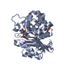
| Related structure data |  6k07C 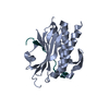 3abdS S: Starting model for refinement C: citing same article ( |
|---|---|
| Similar structure data |


-Links
 PDBj
PDBj


- Assembly
| Deposited unit | 
| ||||||||
|---|---|---|---|---|---|---|---|---|---|
| 1 |
| ||||||||
| Unit cell |
|
-Components
| #1: Protein | Mass: 25239.197 Da / Num. of mol.: 1 / Mutation: R124A,A135D Source method: isolated from a genetically manipulated source Source: (gene. exp.) Homo sapiens (human) / Gene: MAD2L2, MAD2B, REV7 / Production host:  Escherichia coli K-12 (bacteria) / Strain (production host): K-12 / References: UniProt: Q9UI95 Escherichia coli K-12 (bacteria) / Strain (production host): K-12 / References: UniProt: Q9UI95 |
|---|---|
| #2: Protein/peptide | Mass: 3358.924 Da / Num. of mol.: 1 Source method: isolated from a genetically manipulated source Source: (gene. exp.) Homo sapiens (human) / Gene: SHLD3, FLJ26957, RINN1 / Production host: Escherichia coli K-12 (bacteria) / Strain (production host): K-12 / References: UniProt: Q6ZNX1 |
| #3: Chemical | ChemComp-SO4 / Sulfate  Mass: 96.063 Da / Num. of mol.: 1 / Source method: obtained synthetically / Formula: SO4 Mass: 96.063 Da / Num. of mol.: 1 / Source method: obtained synthetically / Formula: SO4 |
| #4: Water | ChemComp-HOH / Water Mass: 18.015 Da / Num. of mol.: 14 / Source method: isolated from a natural source / Formula: H2O Mass: 18.015 Da / Num. of mol.: 14 / Source method: isolated from a natural source / Formula: H2O |
-Experimental details
-Experiment
| Experiment | Method: X-RAY DIFFRACTION / Number of used crystals: 1 |
|---|
- Sample preparation
| Crystal | Density Matthews: 2.49 Å3/Da / Density % sol: 50.72 % |
|---|---|
| Crystal grow | Temperature: 289 K / Method: vapor diffusion, hanging drop / Details: sodium citrate tribasic, ammonium sulfate |
-Data collection
| Diffraction | Mean temperature: 80 K / Serial crystal experiment: N |
|---|---|
| Diffraction source | Source: SYNCHROTRON / Site: SSRF / Beamline: BL19U1 / Wavelength: 0.987 Å |
| Detector | Type: DECTRIS PILATUS 6M-F / Detector: PIXEL / Date: Dec 3, 2018 |
| Radiation | Protocol: SINGLE WAVELENGTH / Monochromatic (M) / Laue (L): M / Scattering type: x-ray |
| Radiation wavelength | Wavelength: 0.987 Å / Relative weight: 1 |
| Reflection | Resolution: 2.31→44.49 Å / Num. obs: 13088 / % possible obs: 99.9 % / Redundancy: 9.3 % / Rpim(I) all: 0.025 / Net I/σ(I): 16.7 |
| Reflection shell | Resolution: 2.31→2.39 Å / Redundancy: 9.8 % / Mean I/σ(I) obs: 2.4 / Num. unique obs: 1283 / Rpim(I) all: 0.453 / % possible all: 99.9 |
- Processing
| Software |
| |||||||||||||||||||||||||||||||||||||||||||||||||||||||||||||||||||||||||||
|---|---|---|---|---|---|---|---|---|---|---|---|---|---|---|---|---|---|---|---|---|---|---|---|---|---|---|---|---|---|---|---|---|---|---|---|---|---|---|---|---|---|---|---|---|---|---|---|---|---|---|---|---|---|---|---|---|---|---|---|---|---|---|---|---|---|---|---|---|---|---|---|---|---|---|---|---|
| Refinement | Method to determine structure: MOLECULAR REPLACEMENT Starting model: 3ABD Resolution: 2.312→44.493 Å / Cross valid method: FREE R-VALUE / σ(F): 1.35
| |||||||||||||||||||||||||||||||||||||||||||||||||||||||||||||||||||||||||||
| Solvent computation | Shrinkage radii: 0.9 Å / VDW probe radii: 1.11 Å | |||||||||||||||||||||||||||||||||||||||||||||||||||||||||||||||||||||||||||
| Refinement step | Cycle: LAST / Resolution: 2.312→44.493 Å
| |||||||||||||||||||||||||||||||||||||||||||||||||||||||||||||||||||||||||||
| Refine LS restraints |
| |||||||||||||||||||||||||||||||||||||||||||||||||||||||||||||||||||||||||||
| LS refinement shell |
| |||||||||||||||||||||||||||||||||||||||||||||||||||||||||||||||||||||||||||
| Refinement TLS params. | Method: refined / Refine-ID: X-RAY DIFFRACTION
| |||||||||||||||||||||||||||||||||||||||||||||||||||||||||||||||||||||||||||
| Refinement TLS group |
|
