 Movie
Movie Controller

[English] 日本語
 Yorodumi
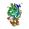
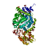
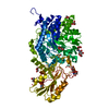
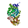
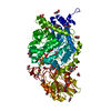
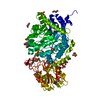
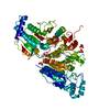
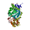
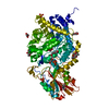
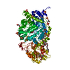
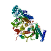
- PDB-6jge: Crystal structure of barley exohydrolaseI W434A mutant in complex... -
+ Open data

- Basic information
| Entry | Database: PDB / ID: 6jge | ||||||
|---|---|---|---|---|---|---|---|
| Title | Crystal structure of barley exohydrolaseI W434A mutant in complex with methyl 2-thio-beta-sophoroside. | ||||||
 Components Components | BETA-D-GLUCAN GLUCOHYDROLASE ISOENZYME EXO1 | ||||||
 Keywords Keywords |  HYDROLASE / Barley exohydrolaseI / enzyme function HYDROLASE / Barley exohydrolaseI / enzyme function | ||||||
| Function / homology |  Function and homology informationbeta-glucosidase / hydrolase activity, hydrolyzing O-glycosyl compounds / membrane => GO:0016020 / carbohydrate metabolic process / extracellular region Function and homology informationbeta-glucosidase / hydrolase activity, hydrolyzing O-glycosyl compounds / membrane => GO:0016020 / carbohydrate metabolic process / extracellular regionSimilarity search - Function | ||||||
| Biological species |  Hordeum vulgare subsp. vulgare (domesticated barley) Hordeum vulgare subsp. vulgare (domesticated barley) | ||||||
| Method | X-RAY DIFFRACTION / SYNCHROTRON / MOLECULAR REPLACEMENT / Resolution: 1.92 Å | ||||||
 Authors Authors | Luang, S. / Streltsov, V.A. / Hrmova, M. | ||||||
 Citation Citation | Journal: Nat Commun / Year: 2022 Title: The evolutionary advantage of an aromatic clamp in plant family 3 glycoside exo-hydrolases. Authors: Luang, S. / Fernandez-Luengo, X. / Nin-Hill, A. / Streltsov, V.A. / Schwerdt, J.G. / Alonso-Gil, S. / Ketudat Cairns, J.R. / Pradeau, S. / Fort, S. / Marechal, J.D. / Masgrau, L. / Rovira, C. / Hrmova, M. | ||||||
| History |
|
- Structure visualization
| Structure viewer | Molecule: MolmilJmol/JSmol |
|---|
- Downloads & links
-Download
| PDBx/mmCIF format | 6jge.cif.gz | 255.9 KB | Display | PDBx/mmCIF format |
|---|---|---|---|---|
| PDB format | pdb6jge.ent.gz | 201.5 KB | Display | PDB format |
| PDBx/mmJSON format | 6jge.json.gz | Tree view | PDBx/mmJSON format | |
| Others |  Other downloads Other downloads |
-Validation report
| Arichive directory | https://data.pdbj.org/pub/pdb/validation_reports/jg/6jgeftp://data.pdbj.org/pub/pdb/validation_reports/jg/6jge | HTTPS FTP |
|---|
-Related structure data
| Related structure data | 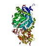 6jg1C  6jg2C 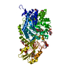 6jg6C  6jg7C  6jgaC  6jgbC  6jgcC  6jgdC 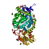 6jggC  6jgkC  6jglC  6jgnC 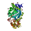 6jgoC 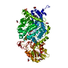 6jgpC  6jgqC  6jgrC 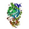 6jgsC 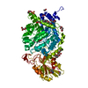 6jgtC 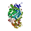 6k6vC 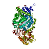 6kufC  6l1jC  6lbbC  6lbvC  6lc5C  3wliS S: Starting model for refinement C: citing same article ( |
|---|---|
| Similar structure data |


-Links
 PDBj
PDBj- Assembly
| Deposited unit | 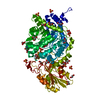
| ||||||||
|---|---|---|---|---|---|---|---|---|---|
| 1 |
| ||||||||
| Unit cell |
|
-Components
-Protein , 1 types, 1 molecules A
| #1: Protein | Mass: 65778.938 Da / Num. of mol.: 1 / Mutation: W434A Source method: isolated from a genetically manipulated source Source: (gene. exp.) Hordeum vulgare subsp. vulgare (domesticated barley)Production host:  Komagataella pastoris (fungus) / References: UniProt: A0A287SCR5, UniProt: Q9XEI3*PLUS Komagataella pastoris (fungus) / References: UniProt: A0A287SCR5, UniProt: Q9XEI3*PLUS |
|---|
-Sugars , 2 types, 2 molecules 
| #2: Polysaccharide | beta-D-glucopyranose-(1-2)-methyl 2-thio-beta-D-glucopyranoside / METHYL 2-THIO-BETA-SOPHOROSIDE  Type: oligosaccharide, Oligosaccharide / Class: Substrate analog / Mass: 372.389 Da / Num. of mol.: 1 Type: oligosaccharide, Oligosaccharide / Class: Substrate analog / Mass: 372.389 Da / Num. of mol.: 1Source method: isolated from a genetically manipulated source Details: oligosaccharide with S-glycosidic bond between monosaccharides References: METHYL 2-THIO-BETA-SOPHOROSIDE |
|---|---|
| #3: Sugar | ChemComp-NAG / N-Acetylglucosamine Type: D-saccharide, beta linking / Mass: 221.208 Da / Num. of mol.: 1 Type: D-saccharide, beta linking / Mass: 221.208 Da / Num. of mol.: 1Source method: isolated from a genetically manipulated source Formula: C8H15NO6 |
-Non-polymers , 4 types, 600 molecules 


| #4: Chemical | ChemComp-GOL / Glycerol Mass: 92.094 Da / Num. of mol.: 15 / Source method: obtained synthetically / Formula: C3H8O3 Mass: 92.094 Da / Num. of mol.: 15 / Source method: obtained synthetically / Formula: C3H8O3#5: Chemical | ChemComp-SO4 / Sulfate Mass: 96.063 Da / Num. of mol.: 8 / Source method: obtained synthetically / Formula: SO4 Mass: 96.063 Da / Num. of mol.: 8 / Source method: obtained synthetically / Formula: SO4#6: Chemical | ChemComp-1PE / | Polyethylene glycol Mass: 238.278 Da / Num. of mol.: 1 / Source method: obtained synthetically / Formula: C10H22O6 / Feature type: SUBJECT OF INVESTIGATION / Comment: precipitant*YM Mass: 238.278 Da / Num. of mol.: 1 / Source method: obtained synthetically / Formula: C10H22O6 / Feature type: SUBJECT OF INVESTIGATION / Comment: precipitant*YM#7: Water | ChemComp-HOH / | WaterMass: 18.015 Da / Num. of mol.: 576 / Source method: isolated from a natural source / Formula: H2O |
|---|
-Details
| Has ligand of interest | Y |
|---|---|
| Sequence details | Amino acid at the position 320 should be Lysine. However, the electron density map is not clear, ...Amino acid at the position 320 should be Lysine. However, the electron density map is not clear, probably side chain of this residue is flexible |
-Experimental details
-Experiment
| Experiment | Method: X-RAY DIFFRACTION / Number of used crystals: 1 |
|---|
- Sample preparation
| Crystal | Density Matthews: 3.5 Å3/Da / Density % sol: 64.82 % |
|---|---|
| Crystal grow | Temperature: 277 K / Method: vapor diffusion, hanging drop / pH: 7 Details: 1.7 M ammonium sulfate, 75 mM HEPES-NaOH buffer, pH 7, containing 7.5 mM sodium acetate and 1.2% (w/v) PEG 400 |
-Data collection
| Diffraction | Mean temperature: 100 K / Serial crystal experiment: N |
|---|---|
| Diffraction source | Source: SYNCHROTRON / Site: Australian Synchrotron  / Beamline: MX1 / Wavelength: 0.9537 Å / Beamline: MX1 / Wavelength: 0.9537 Å |
| Detector | Type: ADSC QUANTUM 210r / Detector: CCD / Date: Nov 22, 2012 / Details: COLLIMATING MIRROR |
| Radiation | Monochromator: DOUBLE-CRYSTAL SI(111) MONOCHROMATOR / Protocol: SINGLE WAVELENGTH / Monochromatic (M) / Laue (L): M / Scattering type: x-ray |
| Radiation wavelength | Wavelength: 0.9537 Å / Relative weight: 1 |
| Reflection | Resolution: 1.92→88.08 Å / Num. obs: 67919 / % possible obs: 99.9 % / Redundancy: 29 % / Rmerge(I) obs: 0.141 / Net I/σ(I): 28.5 |
| Reflection shell | Resolution: 1.92→1.97 Å / Rmerge(I) obs: 0.141 / Num. unique obs: 4913 |
- Processing
| Software |
| ||||||||||||||||||||||||||||||||||||||||||||||||||||||||||||||||||||||||||||||||||||||||||||||||||||
|---|---|---|---|---|---|---|---|---|---|---|---|---|---|---|---|---|---|---|---|---|---|---|---|---|---|---|---|---|---|---|---|---|---|---|---|---|---|---|---|---|---|---|---|---|---|---|---|---|---|---|---|---|---|---|---|---|---|---|---|---|---|---|---|---|---|---|---|---|---|---|---|---|---|---|---|---|---|---|---|---|---|---|---|---|---|---|---|---|---|---|---|---|---|---|---|---|---|---|---|---|---|
| Refinement | Method to determine structure: MOLECULAR REPLACEMENT Starting model: 3WLI Resolution: 1.92→46.1 Å / Cor.coef. Fo:Fc: 0.968 / Cor.coef. Fo:Fc free: 0.959 / SU B: 3.564 / SU ML: 0.055 / Cross valid method: THROUGHOUT / σ(F): 0 / ESU R: 0.093 / ESU R Free: 0.088 / Stereochemistry target values: MAXIMUM LIKELIHOOD Details: HYDROGENS HAVE BEEN ADDED IN THE RIDING POSITIONS U VALUES : WITH TLS ADDED
| ||||||||||||||||||||||||||||||||||||||||||||||||||||||||||||||||||||||||||||||||||||||||||||||||||||
| Solvent computation | Ion probe radii: 0.8 Å / Shrinkage radii: 0.8 Å / VDW probe radii: 1.2 Å / Solvent model: BABINET MODEL WITH MASK | ||||||||||||||||||||||||||||||||||||||||||||||||||||||||||||||||||||||||||||||||||||||||||||||||||||
| Displacement parameters | Biso max: 96.1 Å2 / Biso mean: 20.707 Å2 / Biso min: 8.85 Å2
| ||||||||||||||||||||||||||||||||||||||||||||||||||||||||||||||||||||||||||||||||||||||||||||||||||||
| Refinement step | Cycle: final / Resolution: 1.92→46.1 Å
| ||||||||||||||||||||||||||||||||||||||||||||||||||||||||||||||||||||||||||||||||||||||||||||||||||||
| Refine LS restraints |
| ||||||||||||||||||||||||||||||||||||||||||||||||||||||||||||||||||||||||||||||||||||||||||||||||||||
| LS refinement shell | Resolution: 1.923→1.973 Å / Rfactor Rfree error: 0 / Total num. of bins used: 20
| ||||||||||||||||||||||||||||||||||||||||||||||||||||||||||||||||||||||||||||||||||||||||||||||||||||
| Refinement TLS params. | Method: refined / Refine-ID: X-RAY DIFFRACTION
| ||||||||||||||||||||||||||||||||||||||||||||||||||||||||||||||||||||||||||||||||||||||||||||||||||||
| Refinement TLS group |
|
