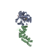
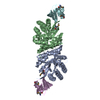
MEMBRANE PROTEIN / mitochondriral fusion / GTPase activity / CMT2A
Function / homology
Function and homology information
: / RHOT2 GTPase cycle / parkin-mediated stimulation of mitophagy in response to mitochondrial depolarization / mitochondrion localization / protein localization to phagophore assembly site / camera-type eye morphogenesis / negative regulation of Ras protein signal transduction / protein targeting to mitochondrion / blastocyst formation / mitochondrial membrane organization ...: / RHOT2 GTPase cycle / parkin-mediated stimulation of mitophagy in response to mitochondrial depolarization / mitochondrion localization / protein localization to phagophore assembly site / camera-type eye morphogenesis / negative regulation of Ras protein signal transduction / protein targeting to mitochondrion / blastocyst formation / mitochondrial membrane organization / mitochondrial fusion / positive regulation of vascular associated smooth muscle cell apoptotic process / response to unfolded protein / aerobic respiration / positive regulation of vascular associated smooth muscle cell proliferation / PINK1-PRKN Mediated Mitophagy / negative regulation of smooth muscle cell proliferation / Hydrolases; Acting on acid anhydrides; Acting on GTP to facilitate cellular and subcellular movement / microtubule cytoskeleton / positive regulation of cold-induced thermogenesis / Factors involved in megakaryocyte development and platelet production / mitochondrial outer membrane / membrane => GO:0016020 / GTPase activity / apoptotic process / ubiquitin protein ligase binding / GTP binding / mitochondrion / cytosol Similarity search - Function
In the structure databanks used in Yorodumi, some data are registered as the other names, "COVID-19 virus" and "2019-nCoV". Here are the details of the virus and the list of structure data.
Jan 31, 2019. EMDB accession codes are about to change! (news from PDBe EMDB page)
EMDB accession codes are about to change! (news from PDBe EMDB page)
The allocation of 4 digits for EMDB accession codes will soon come to an end. Whilst these codes will remain in use, new EMDB accession codes will include an additional digit and will expand incrementally as the available range of codes is exhausted. The current 4-digit format prefixed with “EMD-” (i.e. EMD-XXXX) will advance to a 5-digit format (i.e. EMD-XXXXX), and so on. It is currently estimated that the 4-digit codes will be depleted around Spring 2019, at which point the 5-digit format will come into force.
The EM Navigator/Yorodumi systems omit the EMD- prefix.
Related info.:Q: What is EMD? / ID/Accession-code notation in Yorodumi/EM Navigator
Yorodumi is a browser for structure data from EMDB, PDB, SASBDB, etc.
This page is also the successor to EM Navigator detail page, and also detail information page/front-end page for Omokage search.
The word "yorodu" (or yorozu) is an old Japanese word meaning "ten thousand". "mi" (miru) is to see.
Related info.:EMDB / PDB / SASBDB / Comparison of 3 databanks / Yorodumi Search / Aug 31, 2016. New EM Navigator & Yorodumi / Yorodumi Papers / Jmol/JSmol / Function and homology information / Changes in new EM Navigator and Yorodumi
 Movie
Movie Controller


 Open data
 Basic information
 Components
 Keywords
 MEMBRANE PROTEIN / mitochondriral fusion /
  Function and homology information

 Authors
 China, 1items
  Citation
 Structure visualization
 Downloads & links
 Other downloads
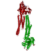


 PDBj
PDBj

 Assembly


 Mass: 59.044 Da / Num. of mol.: 2 / Source method: obtained synthetically / Formula: C2H3O2
 Mass: 40.078 Da / Num. of mol.: 3 / Source method: obtained synthetically / Formula: Ca
 Mass: 18.015 Da / Num. of mol.: 268 / Source method: isolated from a natural source / Formula: H2O
 Sample preparation
 Processing